SlideShare a Scribd company logo
NON NEGATIVE MATRIX FACTORIZATION FOR TUMOR
CLASSIFICATION
Internal Guide
Prof. Kalpesh Jadav
Assistant Prof., EC Department
PIET Limda
Prepared By:
Sahil J Prajapati
M.E. (E.C) 4th
SEM
(130370704517)
OUTLINE
 Motivation
 Introduction
 Challenges
 Types of tumor
 Proposed Method
 Literature survey
 Performance Parameters
 Simulation Results
 Conclusion
 Future Work
 Innovativeness
 References
Motivation:
3
 Brain Tumor is Very Serious Problem
Tumor Detection (Initial Stage)
MRI
BIOPSY
SPINAL TAPE TEST
ANNINOGARM
Disadvantage
 Operative & Painful
 Critical
 Expensive
Ref-http://youtu.be/d95K3unaNC,Cancer.net
Continue…
Present technique includes “Biopsy” procedure which is
operative in manner. Classification based on the imaging
techniques is not acceptable by the radiologist and oncologist
due to required accuracy,because it is difficult to differenciate
between healthy tissue and tumor affected portion.
So, NMF was the technique which motivate in various
applications like face recognition and text mining in terms of
dimensionality and generalization also in image classification.
From this idea the detection of tumor can be made as
Classification of tumor is still a great challenge for radiologists
and neuro-oncologists.
4
Introduction:
Reducing life Expectancy Rate due to Brain Tumor.
Classification and detection of tumors in different medical
images is motivated by the necessity of high accuracy when
dealing with a human life,early detection is best solution for it.
5
Prepare
Data Set
Segmentation
of Tumor
Classification based
on Features
Meningioma
Medullobla-
stroma
Challenges
Automatic classification into one of the five classes such
Astrocytoma, Gliboblastorma Multiforme, Meningioma,
Medulablastroma and Metastatis a challenge.
The treatment of tumors of any types of tumor and its detection
at a primary stage is also a challenge for the Neuro-oncologists
& radiologists in this field.
A highly efficient algorithm to make computation tractable.
Images are not consistent across the dataset due to lighting,
staining, and human preparation variations.
Why tumor classification?
As human brain tumor classification is a difficult task so firstly
understanding the microscopic histology and tumor
morphology by the introduction of microarray technology is
significant and we need to understand its geometry.
Since the resolution of CT and MR images is high so
classification of tumor by various therapies like
radiation,biopsy and chemotherapy has limited to accuracy of
images upto 91.1 to 97%.so classification of tumor by NMF
algorithms have a promising results in improving
accuracy,sensitivity and specfitivity,interpretability.
7
Why Bhattacharyya Coefficient?
Bhattacharyya distance is measure of Similarity of two discrete
or continous probability distributions(Consider two MRI
images),bhattacharyya coefficient is the measure of amount of
overlap betwwn two statistical samples(consider two MRI
images and the measure of dissimilarity of MRI images).
B.C(p,q)=
Here p,q are the two discrete probability distributions points.
Bhattacharya coefficient method is prefered because it easily
locate the target means in the image or frame of interest ,it
finds the dissimilarity between the two image working on
concept of histogram to find the normalize distance,also the
fairity in the image(MRI).
8
1) Astrocytoma
•Astrocytoma can appear in various parts of the brain and
nervous system, including the cerebellum, the cerebrum, the
central areas of the brain, the brainstem, and the spinal cord.
(Grade 2 tumor,infilterative,Slow growing)
9Fig-www.abta.org/astrocytoma.html/
Types of Tumor
2)Medulloblastroma-type of tumor
Medulloblastoma is always located in the cerebellum—the
lower, rear portion of the brain. It is unusual for medullo-
blastomas to spread outside the brain and spinal cord.
10
Fig-www.radiopedia.com/medulloblastroma/html/
3)Meningioma-type of tumor
Meningioma. Although meningiomas are referred to as brain
tumors, they do not grow from brain tissue. They arise from the
meninges, which are three thin layers of tissue covering the brain
and spinal cord. These tumors are most often found near the top
and the outer curve of the brain. Tumors may also form at the
base of the skull.
11Fig-www.abta.org/meningioma/html/
4)Gliboblastroma Multiforme
Gliboblastroma are the generally found in the cerebral
hemisphere of the brain,but can be found any where in the
brain.(Grade 4 tumor,highly infilterative,Rapidly growing
tumor).
12Fig-www.cancer.net/gliboblastroma/html
5) Metastatis
The location of the metastatis brain tumor varies in the brain.
About 85% of metastatic lesions are located in the cerebrum
(the top, largest component of the brain) and 15% are located
in the cerebellum ,the bottom, back part of the brain.(Cancer
Tumor)
13www.abta.org/metastatis/html/
Literature Survey
1. Contour and shape based
2. Region based
3. Machine learning based
4. Statistical based
5. Hybrid and soft computing based
Computer aided diagnosis of human brain tumor through MRI:
A survey and a new algorithm ” EL-sayed A, EL-dahshan Heba M mohsen, kenneth revett, M.saleem, Science Direct.(2014).
Methods of segmentation for detection of brain tumor
This paper mainly on concept of bhattacharyya coefficient which
is used to correlate images by taking the differences in the mirror
images by the concept of correlation and mean square distance.
1.Correlation Images
2.Mean Square Difference
3.Target Localization of the Similarity Images .
4.Bhattacharyya Coefficient
5.Tracking Of the Object
“Bhattacharyya Coefficient In Correlation Of Gray Scale Objects”, M.Sohail
Khalid,M.Umar Ilyas ,Journal Of Multimedia,vol-1,Academy Publisher ,April 2006.
Introduction to bhattacharyya coefficient
This paper discusses about the NMF
1.NMF factorizes a given data matrix into product of two
matrices that contain non negative elements only.
2.Subtraction is forbidden which enhances the sparsity of the
pattern found in data.
3.NMF is feature extraction and to improve upon the linearity
generalization along with improvement in the dimensionality of
the two matrices.
“Non negative matrix factorization and its application to pattern analysis and
text mining”, Jacek M Zurada, Togla Ensari, Ehsaan Hosseini asl, Jan Chorowski, Jacek M Zurada, Togla Ensari, Ehsaan
Hosseini asl, Jan Chorowski,2013 federal conference on computer science and information systems,IEEE,2013
NMF introduction to pattern analysis
This paper discusses about the NMF
1.Image Segmentation And Detection.
2.Histogram Equalization
3.Segmenting Algorithm And Thresholding Operations.
“Brain Tumor Segmentation Using Thresholding,Morphological Operations
And Extraction Of Features Of Tumor”, Deepti Murty S,G..Sadashivappa,2014 international conference ON
Advances In Electronics,Computer And Communication,IEEE 2014.
Brain Tumor Detection and Extraction of
Tumor
This paper discusses about the GLCM And wavelet forms of the
methods with introduction to PNN and ANN network methods
1.Gray Level Co-occurance Matrix(GLCM).
2.Texture Analysis(Feature extraction,Texture
discrimination,texture classification,Shape from texture).
3.Parameters-Energy,Contrast,Entropy,Coorelation coefficient
etc.
4.Classifier like PNN,ANN(used to improve accuracy of the MRI
images )
“MRI Image Classification Using GLCM Texture Features ”, G.Preeti,Mr.Sornagopal,2014
international conference On Signal Processing,IEEE 2014.
Feature Extraction By GLCM With
Comparision to Other Methods
This paper discusses about the Bag Of Model Algorithm
1.Bag Of Words.
2.Texture Feature based on SURF features.
3.Classifier Used .
“An Intergrated Texton And Bag Of Words Classifier For Identifying
Anaplastic Medulloblastroma”, Joseph Galero,Alexender Judkins,David Ellision,Jennifer Baccon,33 International
Conference On the IEEE EMBS,Boston,USA,IEEE 2011.
Brain Tumor Classfication based on Bag Of Model
feature Extraction for Testing And Training Part
This paper discusses Brain Tumor Classification of the Various
Brain Tumor Images By Various Methods
1.Support Vector Machine.
2.Texture And Selection Of features based on PCA,VQ.
3.Classifiers such as ANN,PNN,Neural Networks-classification
also shown based on this Approach.
“MRI Brain Cancer Classification Using Support Vector Machine”, Hari Babu
Nandpura,Dr.S S Salanker,Prof V.R Bora, International Students Conference On Electrical,Electronics And Computer Science,.IEEE
2014.
Brain Tumor Classifiication based By Support
Vector Machine
This paper discusses Brain Tumor Classification of the Various
Brain Tumor Images By Various Methods
1.Confusion Matrix,Accuracy.
2.SVM classifier and it use in creating matrix.
3.Feature Extraction based on GLCM.
“Comparision Of Feature Extraction Techniques To Classify Oral Cancers
Using Image Processing”, K Anuradha,Dr. K Sankaranarayan,International Journal Of Applications and Innovation In
Engineering And Management,Vol-2,Issue-6,June-2013,IJAIEM
Introduction To Confusion Matrix and Accuracy
Why NMF?
NMF is popularized as it tend to decompose images in
meaningful parts that it is able to learn part of the objects.
NMF has enhanced the field of tumor classification but not
only sampling the data but it is used to identify the hidden
features of the tissue or tumor. NMF has improved upon the
singular values decomposition of the variable function to the
multiple function and contained the negative value data that
has not been observed in other ones.its concept is based on
singular values multiplexing of MRI images or can take any
images.
22
Introduction
Non negative matrix factorization is a blind source seperation
techniqe that performs a decomposition of the real valued
matrix of non negative entries into a set of underlying sources
or factors together with their mixing proportions.
It is also used to perform several types of univariate or
multivariate(trade studies across many dimensions) in order to
understand the relationship between variables and their
relevance to the actual problem.they are also used to reduce
hierrarchical “system to system ” dimensionality problem.
23
Mathematical part
Let the value of a pixel in an original input image be V. Let
(WH)iµ be the reconstructed pixel.
If we consider V to be a noisy version of (WH)iµ , then the PDF
of V is
Now we will maximize the log probability of this PDF over W
and H, leaving the relevant objective function to be:
( )
!
))(|(
)(
V
eWH
WHVP
iWHV
i
i
µ
µ
µ =
NMF image formation
Fig-non negative matrix factorization by marshell tappen research scientist,www.cs.ucf.edu/nmf/html/
Proposed Method for Classification
NMF (Nonnegative Matrix Factorization):
Theory: Perception of the whole is based on
perception of its parts.
Comparison with another two matrix factorization
methods:
PCA (Principle Components Analysis)
VQ (Vector quantization )
Compasion-algorithms used before NMF
NMF PCA VQ
Representation: parts- Based holistic holistic
Basis Image: localized feature eigenfaces whole face
Constrains on allow multiple each face is each column of H is
W and H: basis images to approximated by constrained to be a
represent a face,a linear combi- unary vector, every
but only additive nation of all face is approximat-
combinations the eigenfaces ed by a single basis
image.
Workflow
28
MRI dataset images
Image
Preprocessing(contrasting,mean
filtering,Binarization).
Preprocessed image
Detection of tumor (Novel Score Function using
Bhattacharya coefficient,thresholding ,region
growing,otsu’s method approach)
Segment of ROI
feature and Selection(GLCM-feature
Extraction)
Classification of
Tumor(simple,SVM,Baysian Classifier)
Paper-CAD of human brain tumor through MRI:A new algorithm,El-Sayed A,El-Dahshan,Heba A mohsen,expert system
with applications(2014),www.sciencedirect.com
Tumor Segmentation Techniques
29
Tumor Segmentation techniques
Contour
and
shape
based
Region
based
Novel Score
Function
(Bhattacharya
Coefficient)
Watershed
Tumor Classification Techniques
30
Tumor Classification techniques
K- Nearest
Neighbor
Support
Vector
Machine
Artificial
Neural
Network
Texture
Feature
Extraction
Proposed
NMF
Thresholding Approach-Primary approach
towards detection of brain tumor
1st
approach-Thresholding Approach is the simple approach for
the detection of brain tumor but it has one drawback that not all
the 5 tumors are particulary located.
Thresholding Approach is also very helpful to perform the
morphological operation in the tumor image,which help to
carry out the basic operations such as
Preprocessing,Sementation,Classification of the image.
31
Why to go for Novel Score Function for
segmentation?
Region based method include- Preprocessing, Correlation, Post
processing.2nd
approach towards detection of Brain Tumor
It uses Novel Score Function to find dissimilarities in the MRI image,
which gives the detection of tumor accurately.
It gives more accurate results compared to other techniques like
Region based and watershed technique.
32
Region Growing Concept
The drawback of histogram based region detection is that histogram provide
(only the distribution of gray levels),it exploits the important fact that that
pixels which are close together have similar gray value.
 Start with a single seed pixel(seed) and add new pixels slowly.
 Choose the seed pixel.
 Check the neighbouring pixels and add them to the region if they are
similar to seed
 Repeat the above step for the newly added pixels stop if no more pixels are
to be added
 Z-Z(seed)<T.
 Group pixels or sub-regions into larger regions when homogeneity criterion is satisfied
 Region grows around the seed point based on similar properties (grey level, texture, color)
PROS:
 Better in noisy image where edges are hard to identify
CONS:
 Seed point must be specified
 Different seed point will give different results
33
Otsu’s method
It’s the 3rd
approach to detect the brain tumor.
Otsu’s technique is used automatically carry out histogram
shape based image thresholding.
Segmentation is done on basis of a thresholding,due to which
whole image is converted into binary image.
It is used to segment scalar images by generating a binary
partitioning of the image intensities.
Watershed segmentation segregates any image as different
intensity portions and also the tumerous cells have high
proteinous fluid ,which has a high intensity therefore we can
say classify tumors and high intensity tissue of brain.
34
GLCM-Gray level Co-occureence Matrix
GLCM is the method for texture feature extraction proposed by
Haralick in 1998.
Rows and colums are equal to the numbers of gray levels in a
image.which are used in finding the relative frequency
component at the various point to match to the intensity points.
Feature extraction are Contrast,Homogeneity,Entropy,Dis-
similarity,Variance Energy Etc.
GLCM(i,j)=
Where iand J are intensity values of the image and P and Q are
functional Parameters for calculating the frequency betwwen
two pixels.
35
36
Support Vector Machine (SVM)-classifier
intro..
Support vectors
Maximizes
margin
SVMs maximize the margin
around the separating
hyperplane.
 large margin classifiers
The decision function is fully
specified by a subset of training
samples, the support vectors.
Solving SVMs is a quadratic
programming problem
Seen by many as the most
successful current text
classification method *but other discriminative methods
often perform very similarly
Narrower
margin
Why Hyperplane in SVM
Support Vector Machine (SVM) finds an optimal* solution.
Maximizes the distance between the hyperplane and the
“difficult points” close to decision boundary
One intuition: if there are no points near the decision
surface, then there are no very uncertain classification
decisions.
This can be shown by straight line equation Ax+By+C=0.
37
Block Diagram OF SVM(Tumor Classification)
38
Preprocessing OF MRI Dataset
Images
Filtering OF
image(Contrasting,binarization)
Skull Masking(Enhancement etc)
Feature Extraction From Dataset
Feature Selection
SVM Classifier
NO Yes
Normal AbnormalStop
SVM applications
SVM has been used successfully in many real-world problems
- text (and hypertext) categorization
- image classification
- bioinformatics (Protein classification,
Cancer classification)
- hand-written character recognition
39
Classification of Tumor Simple Diagram(Based on
bag of model)
40
Performance parameters
Mean Square Error(MSE)
In Statistics,it measures the average of the square of the errors
that is difference between the estimator and the estimated
value.the difference in value occurs due the randomness or
because the estimator does account for all information that
could produce the more accurate results.
41
Conti…
Peak Signal To Noise Ratio(PSNR)
The phrase peak signal- to-noise ratio, often abbreviated
PSNR, is an engineering term for the ratio between the
maximum possible power of a signal and power of corrupting
noise that affects the fidelity of its representation. Because
many signals have a very wide dynamic range, PSNR is
usually expressed in terms of the log arithmetic decibel scale.
To calculate PSNR value, use the following formula.
42
Conti..
Structural Similarity Idex(SSIM)
The structural similarity (SSIM) index is a method for
measuring the similarity between two images. The SSIM index
is a full reference metric, in other words, the measuring of
image quality based on an initial uncompressed or distortion-
free image as reference. SSIM is designed to improve on
traditional methods like peak signal-to-noise ratio (PSNR) and
mean squared error (MSE), which have proved to be
inconsistent with human eye perception. The SSIM metric is
calculated on various windows of an image. The measure
between two windows x and y of common size N×N is:
43
Simulation Results (Novel Score Function)
Simulation Results-NMF for Tumor detection
45
Performance Parameter for bhattaharya
coefficient+NMF approach
46
GLCM feature extraction
47
Performance On MATLAB-GLCM Feature
extraction
48
Continue…thresholding Approach
49
Fig-Morphological operations Fig-Final output
Performance Parameter for Thresholding
Approach
50
Simulation Results..Region Growing Approach
51
Fig-original image of MRI Fig-Region growing image
Performance Parameter for Region Growing
Approach
52
Simulation results for Otsu’s method
53
Fig-simple morphological operations for MRI image by otsu’s method
Performance Parameter for Otsu’s Method
Approach
54
Classification Results by BOG+Surf
Features(Medulloblastroma Tumor )
55
Classification Results by BOG+Surf
Features(Astrocytoma Tumor )
56
Results Of Classification By SVM-Benign Tumor
57
Results Of Classification By SVM-Malignent
Tumor
58
Calculate the Value Of Accuracy and other
Parameter by Coding NMF+GLCM+SVM For 56
Image Dataset Of all 5tumors
59
Conti……
60
Creation Of Confusion Matrix To find Accuracy-
From the Dataset Of 62 images of all 5 Tumor-
Manual Calculation
61
Comparison Of SVM with existing methods for
Classification
62
Conclusion
Non negative matrix factorization is a tool which is very
helpful for decomposition of images and also for
dimensionality reduction,clustering,segmenting of tumors.
Tumor classification is done to improve upon the
accuracy((95-98%) of the images in such a way that the
segmenting a image give a overall accuracy of the image.
For segmenting the image in set of pixels CAD tools or
machine technology along with region based methods have
shown more accuracy in MRI image that is helpful for neuro-
oncologists in locating the tumor.
Bhattacharya coefficient is the method which is used for
detection(Mirror images) and its results are good in terms of
size and also to compare mirror images .
Future work
In Future work,we will try to classify the to types of Tumors
based on other segmentation algorithms such as SVM+other
algorithms and measure its accuracy and improve upon it .
64
INNOVATIVENESS
Here we propose a combined concept of NMF+Bhattacharyya
coefficient +SVM+GLCM Technique for the detection process
and giving the least errors in the parameter with calculations.
The innovativeness which can be brought to this work is that
the techniques of detection and classification can be combined
and a simple classifier can be used as cascade classifier to get
the results on multiple MRI images and the results can also be
best generated if the work is carried out on DICOM images.
65
REFERENCES
Papers:
1) El-sayed,El-Dashnan,Heba M Mohsen,Kenneth
Revett,M.Saleem,Computer –aided diagonsis of human brain tumor
through MRI:A suvey and a new algorithm”,Expert system with
applications,www.elseiver.com,sciencedirect.com(2014).
2) Rohan kandwal,ashok kumar,An automated system for brain tumor
detection and segmentation”,international journal of advance research in
computer science and software engineering”,www.ijarcsse.com(2014).
3) Tomasz weglinski,Anna Fabijanska,Brain tumor segmentation from MRI
data sets using Region growing approach,MEMStech,polyana-
Svalyava,Ukraine,university of Poland,(2011).
4) Jacek M Zurada,Tolga Ensari,Ehsan Hosenni Asl,Jan Chorwaski,Non
negative matrix factorization and its applications to pattern analysis and
text mining”,2013 federal conference on computer science and information
systems,IEEE(2013).
66
CONTINUE…
5 Jainy Sachdeva,Vinod kumar,indra gupta,Niranjan khandelwal,chirag
kamal Ahuja,Multiclass Brain tumor Classification using GA-SVM,2011
development in E-system Engineering,IEEE(2011).
6 Pawel Szwarc,Aleksander Sobotnicki,Applications of region growing to
brain tumor segmentation”-results,Journal of medical informatics and
technologies,institute (2013).
7 Hui Zhang,Jason E Fritts,Sally Goldman,Image Segmentation
methods:A survey of unsupervised methods,computer vision and image
understanding,www.elseiver.com(sciencedirect.com),(2007).
8 “Deji lu,Yu-Sun,Suiren Wan,Brain tumor classification using Non
negative and Local Non negative matrix factorization”,medical
electronics laboratory,China,IEEE(2013)
9 P.k Srimani,Shahnti Mahesh,Multiclass Tumor classification by using
SVM classifiers”,www.Aijstem.com(2013)
10 Natarajan P,Krishnan N,Natasha Sandeep Kenke,Bhuvanesh Pratap
Singh,”Tumor detection using threshold operation in MRI Brain
Images”,2012 IEEE international conference on computer applications
and research
Continue…
11) A.Jeevitha,P.Narendran,Brain Tumor Segmentation Based On Otsu’s
Thresholding”,International Journal Of Computer Science,Vol-2,Issue-2
February-2013.PAPERIX –Indian Journal Of Research.
12)Deepthi Murthy T,G.Sadashivappa,Brain Tumor Segmentation Using
Thresholding,Morphological Operations And Extraction Of Features Of
Tumor’”,2014 International Conference On Advance In electronicsAnd
Communication,IEEE-2014.
13)M.Sohail Khalid,M.Umar Ilyas,Bhattacharya Coefficient In Correlation Of
Gray Scale Objects”,Journal Of Multimedia,IEEE-2006.
14)Nitish Zulphe,Vrushasen Pawar,GLCM Texture Feature For Brain Tumor
Classification”,International Journal Of Computer Science,Vol-9,Issue-
3,May-2013.
15)A.R Kavitha,Mrs.Kavin Rupa,An Efficient Approach For Brain Tumor
Detection Based On Modified Region Growing In MRI Images,2012
International Conference On Computing Electronics And Electrical
Technologies,IEEE-2012.
68
Non negative matrix factorization ofr tuor classification
Ad

More Related Content

What's hot (20)

PPT on BRAIN TUMOR detection in MRI images based on IMAGE SEGMENTATION
PPT on BRAIN TUMOR detection in MRI images based on  IMAGE SEGMENTATION PPT on BRAIN TUMOR detection in MRI images based on  IMAGE SEGMENTATION
PPT on BRAIN TUMOR detection in MRI images based on IMAGE SEGMENTATION
khanam22
 
An overview of automatic brain tumor detection frommagnetic resonance images
An overview of automatic brain tumor detection frommagnetic resonance imagesAn overview of automatic brain tumor detection frommagnetic resonance images
An overview of automatic brain tumor detection frommagnetic resonance images
Mangesh Lingampalle
 
Brain tumor detection by scanning MRI images (using filtering techniques)
Brain tumor detection by scanning MRI images (using filtering techniques)Brain tumor detection by scanning MRI images (using filtering techniques)
Brain tumor detection by scanning MRI images (using filtering techniques)
Vivek reddy
 
BRAIN TUMOR MRI IMAGE SEGMENTATION AND DETECTION IN IMAGE PROCESSING
BRAIN TUMOR MRI IMAGE SEGMENTATION AND DETECTION IN IMAGE PROCESSINGBRAIN TUMOR MRI IMAGE SEGMENTATION AND DETECTION IN IMAGE PROCESSING
BRAIN TUMOR MRI IMAGE SEGMENTATION AND DETECTION IN IMAGE PROCESSING
Dharshika Shreeganesh
 
Comparitive study of brain tumor detection using morphological operators
Comparitive study of brain tumor detection using morphological operatorsComparitive study of brain tumor detection using morphological operators
Comparitive study of brain tumor detection using morphological operators
eSAT Journals
 
An Image Segmentation and Classification for Brain Tumor Detection using Pill...
An Image Segmentation and Classification for Brain Tumor Detection using Pill...An Image Segmentation and Classification for Brain Tumor Detection using Pill...
An Image Segmentation and Classification for Brain Tumor Detection using Pill...
Association of Scientists, Developers and Faculties
 
B04530612
B04530612B04530612
B04530612
IOSR-JEN
 
Mri brain image segmentatin and classification by modified fcm &amp;svm akorithm
Mri brain image segmentatin and classification by modified fcm &amp;svm akorithmMri brain image segmentatin and classification by modified fcm &amp;svm akorithm
Mri brain image segmentatin and classification by modified fcm &amp;svm akorithm
eSAT Journals
 
Ieee brain tumor
Ieee brain tumorIeee brain tumor
Ieee brain tumor
Prakash Mhaske
 
Brain tumor classification using artificial neural network on mri images
Brain tumor classification using artificial neural network on mri imagesBrain tumor classification using artificial neural network on mri images
Brain tumor classification using artificial neural network on mri images
eSAT Journals
 
IRJET- Brain Tumor Detection using Image Processing and MATLAB Application
IRJET-  	  Brain Tumor Detection using Image Processing and MATLAB ApplicationIRJET-  	  Brain Tumor Detection using Image Processing and MATLAB Application
IRJET- Brain Tumor Detection using Image Processing and MATLAB Application
IRJET Journal
 
Brain Tumor Detection using CNN
Brain Tumor Detection using CNNBrain Tumor Detection using CNN
Brain Tumor Detection using CNN
MohammadRakib8
 
Application of-image-segmentation-in-brain-tumor-detection
Application of-image-segmentation-in-brain-tumor-detectionApplication of-image-segmentation-in-brain-tumor-detection
Application of-image-segmentation-in-brain-tumor-detection
Myat Myint Zu Thin
 
Brain tumor detection using image segmentation ppt
Brain tumor detection using image segmentation pptBrain tumor detection using image segmentation ppt
Brain tumor detection using image segmentation ppt
Roshini Vijayakumar
 
Brain tumor detection and segmentation using watershed segmentation and morph...
Brain tumor detection and segmentation using watershed segmentation and morph...Brain tumor detection and segmentation using watershed segmentation and morph...
Brain tumor detection and segmentation using watershed segmentation and morph...
eSAT Publishing House
 
Brain tumour segmentation based on local independent projection based classif...
Brain tumour segmentation based on local independent projection based classif...Brain tumour segmentation based on local independent projection based classif...
Brain tumour segmentation based on local independent projection based classif...
eSAT Journals
 
BRAIN TUMOR CLASSIFICATION IN 3D-MRI USING FEATURES FROM RADIOMICS AND 3D-CNN...
BRAIN TUMOR CLASSIFICATION IN 3D-MRI USING FEATURES FROM RADIOMICS AND 3D-CNN...BRAIN TUMOR CLASSIFICATION IN 3D-MRI USING FEATURES FROM RADIOMICS AND 3D-CNN...
BRAIN TUMOR CLASSIFICATION IN 3D-MRI USING FEATURES FROM RADIOMICS AND 3D-CNN...
IAEME Publication
 
Brain tumor detection and segmentation using watershed segmentation and morph...
Brain tumor detection and segmentation using watershed segmentation and morph...Brain tumor detection and segmentation using watershed segmentation and morph...
Brain tumor detection and segmentation using watershed segmentation and morph...
eSAT Journals
 
IRJET - Classification of Cancer Images using Deep Learning
IRJET -  	  Classification of Cancer Images using Deep LearningIRJET -  	  Classification of Cancer Images using Deep Learning
IRJET - Classification of Cancer Images using Deep Learning
IRJET Journal
 
Techniques of Brain Cancer Detection from MRI using Machine Learning
Techniques of Brain Cancer Detection from MRI using Machine LearningTechniques of Brain Cancer Detection from MRI using Machine Learning
Techniques of Brain Cancer Detection from MRI using Machine Learning
IRJET Journal
 
PPT on BRAIN TUMOR detection in MRI images based on IMAGE SEGMENTATION
PPT on BRAIN TUMOR detection in MRI images based on  IMAGE SEGMENTATION PPT on BRAIN TUMOR detection in MRI images based on  IMAGE SEGMENTATION
PPT on BRAIN TUMOR detection in MRI images based on IMAGE SEGMENTATION
khanam22
 
An overview of automatic brain tumor detection frommagnetic resonance images
An overview of automatic brain tumor detection frommagnetic resonance imagesAn overview of automatic brain tumor detection frommagnetic resonance images
An overview of automatic brain tumor detection frommagnetic resonance images
Mangesh Lingampalle
 
Brain tumor detection by scanning MRI images (using filtering techniques)
Brain tumor detection by scanning MRI images (using filtering techniques)Brain tumor detection by scanning MRI images (using filtering techniques)
Brain tumor detection by scanning MRI images (using filtering techniques)
Vivek reddy
 
BRAIN TUMOR MRI IMAGE SEGMENTATION AND DETECTION IN IMAGE PROCESSING
BRAIN TUMOR MRI IMAGE SEGMENTATION AND DETECTION IN IMAGE PROCESSINGBRAIN TUMOR MRI IMAGE SEGMENTATION AND DETECTION IN IMAGE PROCESSING
BRAIN TUMOR MRI IMAGE SEGMENTATION AND DETECTION IN IMAGE PROCESSING
Dharshika Shreeganesh
 
Comparitive study of brain tumor detection using morphological operators
Comparitive study of brain tumor detection using morphological operatorsComparitive study of brain tumor detection using morphological operators
Comparitive study of brain tumor detection using morphological operators
eSAT Journals
 
Mri brain image segmentatin and classification by modified fcm &amp;svm akorithm
Mri brain image segmentatin and classification by modified fcm &amp;svm akorithmMri brain image segmentatin and classification by modified fcm &amp;svm akorithm
Mri brain image segmentatin and classification by modified fcm &amp;svm akorithm
eSAT Journals
 
Brain tumor classification using artificial neural network on mri images
Brain tumor classification using artificial neural network on mri imagesBrain tumor classification using artificial neural network on mri images
Brain tumor classification using artificial neural network on mri images
eSAT Journals
 
IRJET- Brain Tumor Detection using Image Processing and MATLAB Application
IRJET-  	  Brain Tumor Detection using Image Processing and MATLAB ApplicationIRJET-  	  Brain Tumor Detection using Image Processing and MATLAB Application
IRJET- Brain Tumor Detection using Image Processing and MATLAB Application
IRJET Journal
 
Brain Tumor Detection using CNN
Brain Tumor Detection using CNNBrain Tumor Detection using CNN
Brain Tumor Detection using CNN
MohammadRakib8
 
Application of-image-segmentation-in-brain-tumor-detection
Application of-image-segmentation-in-brain-tumor-detectionApplication of-image-segmentation-in-brain-tumor-detection
Application of-image-segmentation-in-brain-tumor-detection
Myat Myint Zu Thin
 
Brain tumor detection using image segmentation ppt
Brain tumor detection using image segmentation pptBrain tumor detection using image segmentation ppt
Brain tumor detection using image segmentation ppt
Roshini Vijayakumar
 
Brain tumor detection and segmentation using watershed segmentation and morph...
Brain tumor detection and segmentation using watershed segmentation and morph...Brain tumor detection and segmentation using watershed segmentation and morph...
Brain tumor detection and segmentation using watershed segmentation and morph...
eSAT Publishing House
 
Brain tumour segmentation based on local independent projection based classif...
Brain tumour segmentation based on local independent projection based classif...Brain tumour segmentation based on local independent projection based classif...
Brain tumour segmentation based on local independent projection based classif...
eSAT Journals
 
BRAIN TUMOR CLASSIFICATION IN 3D-MRI USING FEATURES FROM RADIOMICS AND 3D-CNN...
BRAIN TUMOR CLASSIFICATION IN 3D-MRI USING FEATURES FROM RADIOMICS AND 3D-CNN...BRAIN TUMOR CLASSIFICATION IN 3D-MRI USING FEATURES FROM RADIOMICS AND 3D-CNN...
BRAIN TUMOR CLASSIFICATION IN 3D-MRI USING FEATURES FROM RADIOMICS AND 3D-CNN...
IAEME Publication
 
Brain tumor detection and segmentation using watershed segmentation and morph...
Brain tumor detection and segmentation using watershed segmentation and morph...Brain tumor detection and segmentation using watershed segmentation and morph...
Brain tumor detection and segmentation using watershed segmentation and morph...
eSAT Journals
 
IRJET - Classification of Cancer Images using Deep Learning
IRJET -  	  Classification of Cancer Images using Deep LearningIRJET -  	  Classification of Cancer Images using Deep Learning
IRJET - Classification of Cancer Images using Deep Learning
IRJET Journal
 
Techniques of Brain Cancer Detection from MRI using Machine Learning
Techniques of Brain Cancer Detection from MRI using Machine LearningTechniques of Brain Cancer Detection from MRI using Machine Learning
Techniques of Brain Cancer Detection from MRI using Machine Learning
IRJET Journal
 

Similar to Non negative matrix factorization ofr tuor classification (20)

A Review on Brain Disorder Segmentation in MR Images
A Review on Brain Disorder Segmentation in MR ImagesA Review on Brain Disorder Segmentation in MR Images
A Review on Brain Disorder Segmentation in MR Images
IJMER
 
Neural_Network_Based_Brain_Tumor_Detection_Using_Wireless_Infrared_Imaging_Se...
Neural_Network_Based_Brain_Tumor_Detection_Using_Wireless_Infrared_Imaging_Se...Neural_Network_Based_Brain_Tumor_Detection_Using_Wireless_Infrared_Imaging_Se...
Neural_Network_Based_Brain_Tumor_Detection_Using_Wireless_Infrared_Imaging_Se...
RajeshLucky4
 
Bt36430432
Bt36430432Bt36430432
Bt36430432
IJERA Editor
 
A noval methodology for tumor detection in mri images
A noval methodology for tumor detection in mri imagesA noval methodology for tumor detection in mri images
A noval methodology for tumor detection in mri images
DanishAmin26
 
E0413024026
E0413024026E0413024026
E0413024026
ijceronline
 
Paper id 25201482
Paper id 25201482Paper id 25201482
Paper id 25201482
IJRAT
 
L045047880
L045047880L045047880
L045047880
IJERA Editor
 
IRJET- A Novel Segmentation Technique for MRI Brain Tumor Images
IRJET- A Novel Segmentation Technique for MRI Brain Tumor ImagesIRJET- A Novel Segmentation Technique for MRI Brain Tumor Images
IRJET- A Novel Segmentation Technique for MRI Brain Tumor Images
IRJET Journal
 
Ijetcas14 313
Ijetcas14 313Ijetcas14 313
Ijetcas14 313
Iasir Journals
 
BRAIN TUMOR DETECTION
BRAIN TUMOR DETECTIONBRAIN TUMOR DETECTION
BRAIN TUMOR DETECTION
IRJET Journal
 
Survey of various methods used for integrating machine learning into brain tu...
Survey of various methods used for integrating machine learning into brain tu...Survey of various methods used for integrating machine learning into brain tu...
Survey of various methods used for integrating machine learning into brain tu...
Drjabez
 
A Survey On Brain Tumor Detection Techniques
A Survey On Brain Tumor Detection TechniquesA Survey On Brain Tumor Detection Techniques
A Survey On Brain Tumor Detection Techniques
IRJET Journal
 
Paper id 25201472
Paper id 25201472Paper id 25201472
Paper id 25201472
IJRAT
 
IRJET- Brain Tumor Detection using Convolutional Neural Network
IRJET- Brain Tumor Detection using Convolutional Neural NetworkIRJET- Brain Tumor Detection using Convolutional Neural Network
IRJET- Brain Tumor Detection using Convolutional Neural Network
IRJET Journal
 
Brain Tumor Detection System for MRI Image
Brain Tumor Detection System for MRI ImageBrain Tumor Detection System for MRI Image
Brain Tumor Detection System for MRI Image
ijtsrd
 
IRJET- A Study on Brain Tumor Detection Algorithms for MRI Images
IRJET- A Study on Brain Tumor Detection Algorithms for MRI ImagesIRJET- A Study on Brain Tumor Detection Algorithms for MRI Images
IRJET- A Study on Brain Tumor Detection Algorithms for MRI Images
IRJET Journal
 
3D Segmentation of Brain Tumor Imaging
3D Segmentation of Brain Tumor Imaging3D Segmentation of Brain Tumor Imaging
3D Segmentation of Brain Tumor Imaging
IJAEMSJORNAL
 
Automated Intracranial Neoplasm Detection Using Convolutional Neural Networks
Automated Intracranial Neoplasm Detection Using Convolutional Neural NetworksAutomated Intracranial Neoplasm Detection Using Convolutional Neural Networks
Automated Intracranial Neoplasm Detection Using Convolutional Neural Networks
IRJET Journal
 
Optimizing Problem of Brain Tumor Detection using Image Processing
Optimizing Problem of Brain Tumor Detection using Image ProcessingOptimizing Problem of Brain Tumor Detection using Image Processing
Optimizing Problem of Brain Tumor Detection using Image Processing
IRJET Journal
 
BRAIN TUMOR DETECTION USING CNN & ML TECHNIQUES
BRAIN TUMOR DETECTION USING CNN & ML TECHNIQUESBRAIN TUMOR DETECTION USING CNN & ML TECHNIQUES
BRAIN TUMOR DETECTION USING CNN & ML TECHNIQUES
IRJET Journal
 
A Review on Brain Disorder Segmentation in MR Images
A Review on Brain Disorder Segmentation in MR ImagesA Review on Brain Disorder Segmentation in MR Images
A Review on Brain Disorder Segmentation in MR Images
IJMER
 
Neural_Network_Based_Brain_Tumor_Detection_Using_Wireless_Infrared_Imaging_Se...
Neural_Network_Based_Brain_Tumor_Detection_Using_Wireless_Infrared_Imaging_Se...Neural_Network_Based_Brain_Tumor_Detection_Using_Wireless_Infrared_Imaging_Se...
Neural_Network_Based_Brain_Tumor_Detection_Using_Wireless_Infrared_Imaging_Se...
RajeshLucky4
 
A noval methodology for tumor detection in mri images
A noval methodology for tumor detection in mri imagesA noval methodology for tumor detection in mri images
A noval methodology for tumor detection in mri images
DanishAmin26
 
Paper id 25201482
Paper id 25201482Paper id 25201482
Paper id 25201482
IJRAT
 
IRJET- A Novel Segmentation Technique for MRI Brain Tumor Images
IRJET- A Novel Segmentation Technique for MRI Brain Tumor ImagesIRJET- A Novel Segmentation Technique for MRI Brain Tumor Images
IRJET- A Novel Segmentation Technique for MRI Brain Tumor Images
IRJET Journal
 
BRAIN TUMOR DETECTION
BRAIN TUMOR DETECTIONBRAIN TUMOR DETECTION
BRAIN TUMOR DETECTION
IRJET Journal
 
Survey of various methods used for integrating machine learning into brain tu...
Survey of various methods used for integrating machine learning into brain tu...Survey of various methods used for integrating machine learning into brain tu...
Survey of various methods used for integrating machine learning into brain tu...
Drjabez
 
A Survey On Brain Tumor Detection Techniques
A Survey On Brain Tumor Detection TechniquesA Survey On Brain Tumor Detection Techniques
A Survey On Brain Tumor Detection Techniques
IRJET Journal
 
Paper id 25201472
Paper id 25201472Paper id 25201472
Paper id 25201472
IJRAT
 
IRJET- Brain Tumor Detection using Convolutional Neural Network
IRJET- Brain Tumor Detection using Convolutional Neural NetworkIRJET- Brain Tumor Detection using Convolutional Neural Network
IRJET- Brain Tumor Detection using Convolutional Neural Network
IRJET Journal
 
Brain Tumor Detection System for MRI Image
Brain Tumor Detection System for MRI ImageBrain Tumor Detection System for MRI Image
Brain Tumor Detection System for MRI Image
ijtsrd
 
IRJET- A Study on Brain Tumor Detection Algorithms for MRI Images
IRJET- A Study on Brain Tumor Detection Algorithms for MRI ImagesIRJET- A Study on Brain Tumor Detection Algorithms for MRI Images
IRJET- A Study on Brain Tumor Detection Algorithms for MRI Images
IRJET Journal
 
3D Segmentation of Brain Tumor Imaging
3D Segmentation of Brain Tumor Imaging3D Segmentation of Brain Tumor Imaging
3D Segmentation of Brain Tumor Imaging
IJAEMSJORNAL
 
Automated Intracranial Neoplasm Detection Using Convolutional Neural Networks
Automated Intracranial Neoplasm Detection Using Convolutional Neural NetworksAutomated Intracranial Neoplasm Detection Using Convolutional Neural Networks
Automated Intracranial Neoplasm Detection Using Convolutional Neural Networks
IRJET Journal
 
Optimizing Problem of Brain Tumor Detection using Image Processing
Optimizing Problem of Brain Tumor Detection using Image ProcessingOptimizing Problem of Brain Tumor Detection using Image Processing
Optimizing Problem of Brain Tumor Detection using Image Processing
IRJET Journal
 
BRAIN TUMOR DETECTION USING CNN & ML TECHNIQUES
BRAIN TUMOR DETECTION USING CNN & ML TECHNIQUESBRAIN TUMOR DETECTION USING CNN & ML TECHNIQUES
BRAIN TUMOR DETECTION USING CNN & ML TECHNIQUES
IRJET Journal
 
Ad

Recently uploaded (20)

dynastic art of the Pallava dynasty south India
dynastic art of the Pallava dynasty south Indiadynastic art of the Pallava dynasty south India
dynastic art of the Pallava dynasty south India
PrachiSontakke5
 
Presentation of the MIPLM subject matter expert Erdem Kaya
Presentation of the MIPLM subject matter expert Erdem KayaPresentation of the MIPLM subject matter expert Erdem Kaya
Presentation of the MIPLM subject matter expert Erdem Kaya
MIPLM
 
SCI BIZ TECH QUIZ (OPEN) PRELIMS XTASY 2025.pptx
SCI BIZ TECH QUIZ (OPEN) PRELIMS XTASY 2025.pptxSCI BIZ TECH QUIZ (OPEN) PRELIMS XTASY 2025.pptx
SCI BIZ TECH QUIZ (OPEN) PRELIMS XTASY 2025.pptx
Ronisha Das
 
Exercise Physiology MCQS By DR. NASIR MUSTAFA
Exercise Physiology MCQS By DR. NASIR MUSTAFAExercise Physiology MCQS By DR. NASIR MUSTAFA
Exercise Physiology MCQS By DR. NASIR MUSTAFA
Dr. Nasir Mustafa
 
To study the nervous system of insect.pptx
To study the nervous system of insect.pptxTo study the nervous system of insect.pptx
To study the nervous system of insect.pptx
Arshad Shaikh
 
Presentation on Tourism Product Development By Md Shaifullar Rabbi
Presentation on Tourism Product Development By Md Shaifullar RabbiPresentation on Tourism Product Development By Md Shaifullar Rabbi
Presentation on Tourism Product Development By Md Shaifullar Rabbi
Md Shaifullar Rabbi
 
Political History of Pala dynasty Pala Rulers NEP.pptx
Political History of Pala dynasty Pala Rulers NEP.pptxPolitical History of Pala dynasty Pala Rulers NEP.pptx
Political History of Pala dynasty Pala Rulers NEP.pptx
Arya Mahila P. G. College, Banaras Hindu University, Varanasi, India.
 
World war-1(Causes & impacts at a glance) PPT by Simanchala Sarab(BABed,sem-4...
World war-1(Causes & impacts at a glance) PPT by Simanchala Sarab(BABed,sem-4...World war-1(Causes & impacts at a glance) PPT by Simanchala Sarab(BABed,sem-4...
World war-1(Causes & impacts at a glance) PPT by Simanchala Sarab(BABed,sem-4...
larencebapu132
 
Introduction to Vibe Coding and Vibe Engineering
Introduction to Vibe Coding and Vibe EngineeringIntroduction to Vibe Coding and Vibe Engineering
Introduction to Vibe Coding and Vibe Engineering
Damian T. Gordon
 
How to track Cost and Revenue using Analytic Accounts in odoo Accounting, App...
How to track Cost and Revenue using Analytic Accounts in odoo Accounting, App...How to track Cost and Revenue using Analytic Accounts in odoo Accounting, App...
How to track Cost and Revenue using Analytic Accounts in odoo Accounting, App...
Celine George
 
pulse ppt.pptx Types of pulse , characteristics of pulse , Alteration of pulse
pulse  ppt.pptx Types of pulse , characteristics of pulse , Alteration of pulsepulse  ppt.pptx Types of pulse , characteristics of pulse , Alteration of pulse
pulse ppt.pptx Types of pulse , characteristics of pulse , Alteration of pulse
sushreesangita003
 
Introduction-to-Communication-and-Media-Studies-1736283331.pdf
Introduction-to-Communication-and-Media-Studies-1736283331.pdfIntroduction-to-Communication-and-Media-Studies-1736283331.pdf
Introduction-to-Communication-and-Media-Studies-1736283331.pdf
james5028
 
Metamorphosis: Life's Transformative Journey
Metamorphosis: Life's Transformative JourneyMetamorphosis: Life's Transformative Journey
Metamorphosis: Life's Transformative Journey
Arshad Shaikh
 
How to Set warnings for invoicing specific customers in odoo
How to Set warnings for invoicing specific customers in odooHow to Set warnings for invoicing specific customers in odoo
How to Set warnings for invoicing specific customers in odoo
Celine George
 
Engage Donors Through Powerful Storytelling.pdf
Engage Donors Through Powerful Storytelling.pdfEngage Donors Through Powerful Storytelling.pdf
Engage Donors Through Powerful Storytelling.pdf
TechSoup
 
Operations Management (Dr. Abdulfatah Salem).pdf
Operations Management (Dr. Abdulfatah Salem).pdfOperations Management (Dr. Abdulfatah Salem).pdf
Operations Management (Dr. Abdulfatah Salem).pdf
Arab Academy for Science, Technology and Maritime Transport
 
CBSE - Grade 8 - Science - Chemistry - Metals and Non Metals - Worksheet
CBSE - Grade 8 - Science - Chemistry - Metals and Non Metals - WorksheetCBSE - Grade 8 - Science - Chemistry - Metals and Non Metals - Worksheet
CBSE - Grade 8 - Science - Chemistry - Metals and Non Metals - Worksheet
Sritoma Majumder
 
Biophysics Chapter 3 Methods of Studying Macromolecules.pdf
Biophysics Chapter 3 Methods of Studying Macromolecules.pdfBiophysics Chapter 3 Methods of Studying Macromolecules.pdf
Biophysics Chapter 3 Methods of Studying Macromolecules.pdf
PKLI-Institute of Nursing and Allied Health Sciences Lahore , Pakistan.
 
To study Digestive system of insect.pptx
To study Digestive system of insect.pptxTo study Digestive system of insect.pptx
To study Digestive system of insect.pptx
Arshad Shaikh
 
Grade 2 - Mathematics - Printable Worksheet
Grade 2 - Mathematics - Printable WorksheetGrade 2 - Mathematics - Printable Worksheet
Grade 2 - Mathematics - Printable Worksheet
Sritoma Majumder
 
dynastic art of the Pallava dynasty south India
dynastic art of the Pallava dynasty south Indiadynastic art of the Pallava dynasty south India
dynastic art of the Pallava dynasty south India
PrachiSontakke5
 
Presentation of the MIPLM subject matter expert Erdem Kaya
Presentation of the MIPLM subject matter expert Erdem KayaPresentation of the MIPLM subject matter expert Erdem Kaya
Presentation of the MIPLM subject matter expert Erdem Kaya
MIPLM
 
SCI BIZ TECH QUIZ (OPEN) PRELIMS XTASY 2025.pptx
SCI BIZ TECH QUIZ (OPEN) PRELIMS XTASY 2025.pptxSCI BIZ TECH QUIZ (OPEN) PRELIMS XTASY 2025.pptx
SCI BIZ TECH QUIZ (OPEN) PRELIMS XTASY 2025.pptx
Ronisha Das
 
Exercise Physiology MCQS By DR. NASIR MUSTAFA
Exercise Physiology MCQS By DR. NASIR MUSTAFAExercise Physiology MCQS By DR. NASIR MUSTAFA
Exercise Physiology MCQS By DR. NASIR MUSTAFA
Dr. Nasir Mustafa
 
To study the nervous system of insect.pptx
To study the nervous system of insect.pptxTo study the nervous system of insect.pptx
To study the nervous system of insect.pptx
Arshad Shaikh
 
Presentation on Tourism Product Development By Md Shaifullar Rabbi
Presentation on Tourism Product Development By Md Shaifullar RabbiPresentation on Tourism Product Development By Md Shaifullar Rabbi
Presentation on Tourism Product Development By Md Shaifullar Rabbi
Md Shaifullar Rabbi
 
World war-1(Causes & impacts at a glance) PPT by Simanchala Sarab(BABed,sem-4...
World war-1(Causes & impacts at a glance) PPT by Simanchala Sarab(BABed,sem-4...World war-1(Causes & impacts at a glance) PPT by Simanchala Sarab(BABed,sem-4...
World war-1(Causes & impacts at a glance) PPT by Simanchala Sarab(BABed,sem-4...
larencebapu132
 
Introduction to Vibe Coding and Vibe Engineering
Introduction to Vibe Coding and Vibe EngineeringIntroduction to Vibe Coding and Vibe Engineering
Introduction to Vibe Coding and Vibe Engineering
Damian T. Gordon
 
How to track Cost and Revenue using Analytic Accounts in odoo Accounting, App...
How to track Cost and Revenue using Analytic Accounts in odoo Accounting, App...How to track Cost and Revenue using Analytic Accounts in odoo Accounting, App...
How to track Cost and Revenue using Analytic Accounts in odoo Accounting, App...
Celine George
 
pulse ppt.pptx Types of pulse , characteristics of pulse , Alteration of pulse
pulse  ppt.pptx Types of pulse , characteristics of pulse , Alteration of pulsepulse  ppt.pptx Types of pulse , characteristics of pulse , Alteration of pulse
pulse ppt.pptx Types of pulse , characteristics of pulse , Alteration of pulse
sushreesangita003
 
Introduction-to-Communication-and-Media-Studies-1736283331.pdf
Introduction-to-Communication-and-Media-Studies-1736283331.pdfIntroduction-to-Communication-and-Media-Studies-1736283331.pdf
Introduction-to-Communication-and-Media-Studies-1736283331.pdf
james5028
 
Metamorphosis: Life's Transformative Journey
Metamorphosis: Life's Transformative JourneyMetamorphosis: Life's Transformative Journey
Metamorphosis: Life's Transformative Journey
Arshad Shaikh
 
How to Set warnings for invoicing specific customers in odoo
How to Set warnings for invoicing specific customers in odooHow to Set warnings for invoicing specific customers in odoo
How to Set warnings for invoicing specific customers in odoo
Celine George
 
Engage Donors Through Powerful Storytelling.pdf
Engage Donors Through Powerful Storytelling.pdfEngage Donors Through Powerful Storytelling.pdf
Engage Donors Through Powerful Storytelling.pdf
TechSoup
 
CBSE - Grade 8 - Science - Chemistry - Metals and Non Metals - Worksheet
CBSE - Grade 8 - Science - Chemistry - Metals and Non Metals - WorksheetCBSE - Grade 8 - Science - Chemistry - Metals and Non Metals - Worksheet
CBSE - Grade 8 - Science - Chemistry - Metals and Non Metals - Worksheet
Sritoma Majumder
 
To study Digestive system of insect.pptx
To study Digestive system of insect.pptxTo study Digestive system of insect.pptx
To study Digestive system of insect.pptx
Arshad Shaikh
 
Grade 2 - Mathematics - Printable Worksheet
Grade 2 - Mathematics - Printable WorksheetGrade 2 - Mathematics - Printable Worksheet
Grade 2 - Mathematics - Printable Worksheet
Sritoma Majumder
 
Ad

Non negative matrix factorization ofr tuor classification

  • 1. NON NEGATIVE MATRIX FACTORIZATION FOR TUMOR CLASSIFICATION Internal Guide Prof. Kalpesh Jadav Assistant Prof., EC Department PIET Limda Prepared By: Sahil J Prajapati M.E. (E.C) 4th SEM (130370704517)
  • 2. OUTLINE  Motivation  Introduction  Challenges  Types of tumor  Proposed Method  Literature survey  Performance Parameters  Simulation Results  Conclusion  Future Work  Innovativeness  References
  • 3. Motivation: 3  Brain Tumor is Very Serious Problem Tumor Detection (Initial Stage) MRI BIOPSY SPINAL TAPE TEST ANNINOGARM Disadvantage  Operative & Painful  Critical  Expensive Ref-http://youtu.be/d95K3unaNC,Cancer.net
  • 4. Continue… Present technique includes “Biopsy” procedure which is operative in manner. Classification based on the imaging techniques is not acceptable by the radiologist and oncologist due to required accuracy,because it is difficult to differenciate between healthy tissue and tumor affected portion. So, NMF was the technique which motivate in various applications like face recognition and text mining in terms of dimensionality and generalization also in image classification. From this idea the detection of tumor can be made as Classification of tumor is still a great challenge for radiologists and neuro-oncologists. 4
  • 5. Introduction: Reducing life Expectancy Rate due to Brain Tumor. Classification and detection of tumors in different medical images is motivated by the necessity of high accuracy when dealing with a human life,early detection is best solution for it. 5 Prepare Data Set Segmentation of Tumor Classification based on Features Meningioma Medullobla- stroma
  • 6. Challenges Automatic classification into one of the five classes such Astrocytoma, Gliboblastorma Multiforme, Meningioma, Medulablastroma and Metastatis a challenge. The treatment of tumors of any types of tumor and its detection at a primary stage is also a challenge for the Neuro-oncologists & radiologists in this field. A highly efficient algorithm to make computation tractable. Images are not consistent across the dataset due to lighting, staining, and human preparation variations.
  • 7. Why tumor classification? As human brain tumor classification is a difficult task so firstly understanding the microscopic histology and tumor morphology by the introduction of microarray technology is significant and we need to understand its geometry. Since the resolution of CT and MR images is high so classification of tumor by various therapies like radiation,biopsy and chemotherapy has limited to accuracy of images upto 91.1 to 97%.so classification of tumor by NMF algorithms have a promising results in improving accuracy,sensitivity and specfitivity,interpretability. 7
  • 8. Why Bhattacharyya Coefficient? Bhattacharyya distance is measure of Similarity of two discrete or continous probability distributions(Consider two MRI images),bhattacharyya coefficient is the measure of amount of overlap betwwn two statistical samples(consider two MRI images and the measure of dissimilarity of MRI images). B.C(p,q)= Here p,q are the two discrete probability distributions points. Bhattacharya coefficient method is prefered because it easily locate the target means in the image or frame of interest ,it finds the dissimilarity between the two image working on concept of histogram to find the normalize distance,also the fairity in the image(MRI). 8
  • 9. 1) Astrocytoma •Astrocytoma can appear in various parts of the brain and nervous system, including the cerebellum, the cerebrum, the central areas of the brain, the brainstem, and the spinal cord. (Grade 2 tumor,infilterative,Slow growing) 9Fig-www.abta.org/astrocytoma.html/ Types of Tumor
  • 10. 2)Medulloblastroma-type of tumor Medulloblastoma is always located in the cerebellum—the lower, rear portion of the brain. It is unusual for medullo- blastomas to spread outside the brain and spinal cord. 10 Fig-www.radiopedia.com/medulloblastroma/html/
  • 11. 3)Meningioma-type of tumor Meningioma. Although meningiomas are referred to as brain tumors, they do not grow from brain tissue. They arise from the meninges, which are three thin layers of tissue covering the brain and spinal cord. These tumors are most often found near the top and the outer curve of the brain. Tumors may also form at the base of the skull. 11Fig-www.abta.org/meningioma/html/
  • 12. 4)Gliboblastroma Multiforme Gliboblastroma are the generally found in the cerebral hemisphere of the brain,but can be found any where in the brain.(Grade 4 tumor,highly infilterative,Rapidly growing tumor). 12Fig-www.cancer.net/gliboblastroma/html
  • 13. 5) Metastatis The location of the metastatis brain tumor varies in the brain. About 85% of metastatic lesions are located in the cerebrum (the top, largest component of the brain) and 15% are located in the cerebellum ,the bottom, back part of the brain.(Cancer Tumor) 13www.abta.org/metastatis/html/
  • 14. Literature Survey 1. Contour and shape based 2. Region based 3. Machine learning based 4. Statistical based 5. Hybrid and soft computing based Computer aided diagnosis of human brain tumor through MRI: A survey and a new algorithm ” EL-sayed A, EL-dahshan Heba M mohsen, kenneth revett, M.saleem, Science Direct.(2014). Methods of segmentation for detection of brain tumor
  • 15. This paper mainly on concept of bhattacharyya coefficient which is used to correlate images by taking the differences in the mirror images by the concept of correlation and mean square distance. 1.Correlation Images 2.Mean Square Difference 3.Target Localization of the Similarity Images . 4.Bhattacharyya Coefficient 5.Tracking Of the Object “Bhattacharyya Coefficient In Correlation Of Gray Scale Objects”, M.Sohail Khalid,M.Umar Ilyas ,Journal Of Multimedia,vol-1,Academy Publisher ,April 2006. Introduction to bhattacharyya coefficient
  • 16. This paper discusses about the NMF 1.NMF factorizes a given data matrix into product of two matrices that contain non negative elements only. 2.Subtraction is forbidden which enhances the sparsity of the pattern found in data. 3.NMF is feature extraction and to improve upon the linearity generalization along with improvement in the dimensionality of the two matrices. “Non negative matrix factorization and its application to pattern analysis and text mining”, Jacek M Zurada, Togla Ensari, Ehsaan Hosseini asl, Jan Chorowski, Jacek M Zurada, Togla Ensari, Ehsaan Hosseini asl, Jan Chorowski,2013 federal conference on computer science and information systems,IEEE,2013 NMF introduction to pattern analysis
  • 17. This paper discusses about the NMF 1.Image Segmentation And Detection. 2.Histogram Equalization 3.Segmenting Algorithm And Thresholding Operations. “Brain Tumor Segmentation Using Thresholding,Morphological Operations And Extraction Of Features Of Tumor”, Deepti Murty S,G..Sadashivappa,2014 international conference ON Advances In Electronics,Computer And Communication,IEEE 2014. Brain Tumor Detection and Extraction of Tumor
  • 18. This paper discusses about the GLCM And wavelet forms of the methods with introduction to PNN and ANN network methods 1.Gray Level Co-occurance Matrix(GLCM). 2.Texture Analysis(Feature extraction,Texture discrimination,texture classification,Shape from texture). 3.Parameters-Energy,Contrast,Entropy,Coorelation coefficient etc. 4.Classifier like PNN,ANN(used to improve accuracy of the MRI images ) “MRI Image Classification Using GLCM Texture Features ”, G.Preeti,Mr.Sornagopal,2014 international conference On Signal Processing,IEEE 2014. Feature Extraction By GLCM With Comparision to Other Methods
  • 19. This paper discusses about the Bag Of Model Algorithm 1.Bag Of Words. 2.Texture Feature based on SURF features. 3.Classifier Used . “An Intergrated Texton And Bag Of Words Classifier For Identifying Anaplastic Medulloblastroma”, Joseph Galero,Alexender Judkins,David Ellision,Jennifer Baccon,33 International Conference On the IEEE EMBS,Boston,USA,IEEE 2011. Brain Tumor Classfication based on Bag Of Model feature Extraction for Testing And Training Part
  • 20. This paper discusses Brain Tumor Classification of the Various Brain Tumor Images By Various Methods 1.Support Vector Machine. 2.Texture And Selection Of features based on PCA,VQ. 3.Classifiers such as ANN,PNN,Neural Networks-classification also shown based on this Approach. “MRI Brain Cancer Classification Using Support Vector Machine”, Hari Babu Nandpura,Dr.S S Salanker,Prof V.R Bora, International Students Conference On Electrical,Electronics And Computer Science,.IEEE 2014. Brain Tumor Classifiication based By Support Vector Machine
  • 21. This paper discusses Brain Tumor Classification of the Various Brain Tumor Images By Various Methods 1.Confusion Matrix,Accuracy. 2.SVM classifier and it use in creating matrix. 3.Feature Extraction based on GLCM. “Comparision Of Feature Extraction Techniques To Classify Oral Cancers Using Image Processing”, K Anuradha,Dr. K Sankaranarayan,International Journal Of Applications and Innovation In Engineering And Management,Vol-2,Issue-6,June-2013,IJAIEM Introduction To Confusion Matrix and Accuracy
  • 22. Why NMF? NMF is popularized as it tend to decompose images in meaningful parts that it is able to learn part of the objects. NMF has enhanced the field of tumor classification but not only sampling the data but it is used to identify the hidden features of the tissue or tumor. NMF has improved upon the singular values decomposition of the variable function to the multiple function and contained the negative value data that has not been observed in other ones.its concept is based on singular values multiplexing of MRI images or can take any images. 22
  • 23. Introduction Non negative matrix factorization is a blind source seperation techniqe that performs a decomposition of the real valued matrix of non negative entries into a set of underlying sources or factors together with their mixing proportions. It is also used to perform several types of univariate or multivariate(trade studies across many dimensions) in order to understand the relationship between variables and their relevance to the actual problem.they are also used to reduce hierrarchical “system to system ” dimensionality problem. 23
  • 24. Mathematical part Let the value of a pixel in an original input image be V. Let (WH)iµ be the reconstructed pixel. If we consider V to be a noisy version of (WH)iµ , then the PDF of V is Now we will maximize the log probability of this PDF over W and H, leaving the relevant objective function to be: ( ) ! ))(|( )( V eWH WHVP iWHV i i µ µ µ =
  • 25. NMF image formation Fig-non negative matrix factorization by marshell tappen research scientist,www.cs.ucf.edu/nmf/html/
  • 26. Proposed Method for Classification NMF (Nonnegative Matrix Factorization): Theory: Perception of the whole is based on perception of its parts. Comparison with another two matrix factorization methods: PCA (Principle Components Analysis) VQ (Vector quantization )
  • 27. Compasion-algorithms used before NMF NMF PCA VQ Representation: parts- Based holistic holistic Basis Image: localized feature eigenfaces whole face Constrains on allow multiple each face is each column of H is W and H: basis images to approximated by constrained to be a represent a face,a linear combi- unary vector, every but only additive nation of all face is approximat- combinations the eigenfaces ed by a single basis image.
  • 28. Workflow 28 MRI dataset images Image Preprocessing(contrasting,mean filtering,Binarization). Preprocessed image Detection of tumor (Novel Score Function using Bhattacharya coefficient,thresholding ,region growing,otsu’s method approach) Segment of ROI feature and Selection(GLCM-feature Extraction) Classification of Tumor(simple,SVM,Baysian Classifier) Paper-CAD of human brain tumor through MRI:A new algorithm,El-Sayed A,El-Dahshan,Heba A mohsen,expert system with applications(2014),www.sciencedirect.com
  • 29. Tumor Segmentation Techniques 29 Tumor Segmentation techniques Contour and shape based Region based Novel Score Function (Bhattacharya Coefficient) Watershed
  • 30. Tumor Classification Techniques 30 Tumor Classification techniques K- Nearest Neighbor Support Vector Machine Artificial Neural Network Texture Feature Extraction Proposed NMF
  • 31. Thresholding Approach-Primary approach towards detection of brain tumor 1st approach-Thresholding Approach is the simple approach for the detection of brain tumor but it has one drawback that not all the 5 tumors are particulary located. Thresholding Approach is also very helpful to perform the morphological operation in the tumor image,which help to carry out the basic operations such as Preprocessing,Sementation,Classification of the image. 31
  • 32. Why to go for Novel Score Function for segmentation? Region based method include- Preprocessing, Correlation, Post processing.2nd approach towards detection of Brain Tumor It uses Novel Score Function to find dissimilarities in the MRI image, which gives the detection of tumor accurately. It gives more accurate results compared to other techniques like Region based and watershed technique. 32
  • 33. Region Growing Concept The drawback of histogram based region detection is that histogram provide (only the distribution of gray levels),it exploits the important fact that that pixels which are close together have similar gray value.  Start with a single seed pixel(seed) and add new pixels slowly.  Choose the seed pixel.  Check the neighbouring pixels and add them to the region if they are similar to seed  Repeat the above step for the newly added pixels stop if no more pixels are to be added  Z-Z(seed)<T.  Group pixels or sub-regions into larger regions when homogeneity criterion is satisfied  Region grows around the seed point based on similar properties (grey level, texture, color) PROS:  Better in noisy image where edges are hard to identify CONS:  Seed point must be specified  Different seed point will give different results 33
  • 34. Otsu’s method It’s the 3rd approach to detect the brain tumor. Otsu’s technique is used automatically carry out histogram shape based image thresholding. Segmentation is done on basis of a thresholding,due to which whole image is converted into binary image. It is used to segment scalar images by generating a binary partitioning of the image intensities. Watershed segmentation segregates any image as different intensity portions and also the tumerous cells have high proteinous fluid ,which has a high intensity therefore we can say classify tumors and high intensity tissue of brain. 34
  • 35. GLCM-Gray level Co-occureence Matrix GLCM is the method for texture feature extraction proposed by Haralick in 1998. Rows and colums are equal to the numbers of gray levels in a image.which are used in finding the relative frequency component at the various point to match to the intensity points. Feature extraction are Contrast,Homogeneity,Entropy,Dis- similarity,Variance Energy Etc. GLCM(i,j)= Where iand J are intensity values of the image and P and Q are functional Parameters for calculating the frequency betwwen two pixels. 35
  • 36. 36 Support Vector Machine (SVM)-classifier intro.. Support vectors Maximizes margin SVMs maximize the margin around the separating hyperplane.  large margin classifiers The decision function is fully specified by a subset of training samples, the support vectors. Solving SVMs is a quadratic programming problem Seen by many as the most successful current text classification method *but other discriminative methods often perform very similarly Narrower margin
  • 37. Why Hyperplane in SVM Support Vector Machine (SVM) finds an optimal* solution. Maximizes the distance between the hyperplane and the “difficult points” close to decision boundary One intuition: if there are no points near the decision surface, then there are no very uncertain classification decisions. This can be shown by straight line equation Ax+By+C=0. 37
  • 38. Block Diagram OF SVM(Tumor Classification) 38 Preprocessing OF MRI Dataset Images Filtering OF image(Contrasting,binarization) Skull Masking(Enhancement etc) Feature Extraction From Dataset Feature Selection SVM Classifier NO Yes Normal AbnormalStop
  • 39. SVM applications SVM has been used successfully in many real-world problems - text (and hypertext) categorization - image classification - bioinformatics (Protein classification, Cancer classification) - hand-written character recognition 39
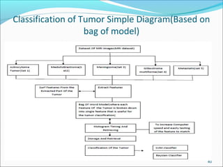
  • 40. Classification of Tumor Simple Diagram(Based on bag of model) 40
  • 41. Performance parameters Mean Square Error(MSE) In Statistics,it measures the average of the square of the errors that is difference between the estimator and the estimated value.the difference in value occurs due the randomness or because the estimator does account for all information that could produce the more accurate results. 41
  • 42. Conti… Peak Signal To Noise Ratio(PSNR) The phrase peak signal- to-noise ratio, often abbreviated PSNR, is an engineering term for the ratio between the maximum possible power of a signal and power of corrupting noise that affects the fidelity of its representation. Because many signals have a very wide dynamic range, PSNR is usually expressed in terms of the log arithmetic decibel scale. To calculate PSNR value, use the following formula. 42
  • 43. Conti.. Structural Similarity Idex(SSIM) The structural similarity (SSIM) index is a method for measuring the similarity between two images. The SSIM index is a full reference metric, in other words, the measuring of image quality based on an initial uncompressed or distortion- free image as reference. SSIM is designed to improve on traditional methods like peak signal-to-noise ratio (PSNR) and mean squared error (MSE), which have proved to be inconsistent with human eye perception. The SSIM metric is calculated on various windows of an image. The measure between two windows x and y of common size N×N is: 43
  • 44. Simulation Results (Novel Score Function)
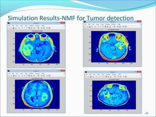
  • 45. Simulation Results-NMF for Tumor detection 45
  • 46. Performance Parameter for bhattaharya coefficient+NMF approach 46
  • 48. Performance On MATLAB-GLCM Feature extraction 48
  • 50. Performance Parameter for Thresholding Approach 50
  • 51. Simulation Results..Region Growing Approach 51 Fig-original image of MRI Fig-Region growing image
  • 52. Performance Parameter for Region Growing Approach 52
  • 53. Simulation results for Otsu’s method 53 Fig-simple morphological operations for MRI image by otsu’s method
  • 54. Performance Parameter for Otsu’s Method Approach 54
  • 55. Classification Results by BOG+Surf Features(Medulloblastroma Tumor ) 55
  • 56. Classification Results by BOG+Surf Features(Astrocytoma Tumor ) 56
  • 57. Results Of Classification By SVM-Benign Tumor 57
  • 58. Results Of Classification By SVM-Malignent Tumor 58
  • 59. Calculate the Value Of Accuracy and other Parameter by Coding NMF+GLCM+SVM For 56 Image Dataset Of all 5tumors 59
  • 61. Creation Of Confusion Matrix To find Accuracy- From the Dataset Of 62 images of all 5 Tumor- Manual Calculation 61
  • 62. Comparison Of SVM with existing methods for Classification 62
  • 63. Conclusion Non negative matrix factorization is a tool which is very helpful for decomposition of images and also for dimensionality reduction,clustering,segmenting of tumors. Tumor classification is done to improve upon the accuracy((95-98%) of the images in such a way that the segmenting a image give a overall accuracy of the image. For segmenting the image in set of pixels CAD tools or machine technology along with region based methods have shown more accuracy in MRI image that is helpful for neuro- oncologists in locating the tumor. Bhattacharya coefficient is the method which is used for detection(Mirror images) and its results are good in terms of size and also to compare mirror images .
  • 64. Future work In Future work,we will try to classify the to types of Tumors based on other segmentation algorithms such as SVM+other algorithms and measure its accuracy and improve upon it . 64
  • 65. INNOVATIVENESS Here we propose a combined concept of NMF+Bhattacharyya coefficient +SVM+GLCM Technique for the detection process and giving the least errors in the parameter with calculations. The innovativeness which can be brought to this work is that the techniques of detection and classification can be combined and a simple classifier can be used as cascade classifier to get the results on multiple MRI images and the results can also be best generated if the work is carried out on DICOM images. 65
  • 66. REFERENCES Papers: 1) El-sayed,El-Dashnan,Heba M Mohsen,Kenneth Revett,M.Saleem,Computer –aided diagonsis of human brain tumor through MRI:A suvey and a new algorithm”,Expert system with applications,www.elseiver.com,sciencedirect.com(2014). 2) Rohan kandwal,ashok kumar,An automated system for brain tumor detection and segmentation”,international journal of advance research in computer science and software engineering”,www.ijarcsse.com(2014). 3) Tomasz weglinski,Anna Fabijanska,Brain tumor segmentation from MRI data sets using Region growing approach,MEMStech,polyana- Svalyava,Ukraine,university of Poland,(2011). 4) Jacek M Zurada,Tolga Ensari,Ehsan Hosenni Asl,Jan Chorwaski,Non negative matrix factorization and its applications to pattern analysis and text mining”,2013 federal conference on computer science and information systems,IEEE(2013). 66
  • 67. CONTINUE… 5 Jainy Sachdeva,Vinod kumar,indra gupta,Niranjan khandelwal,chirag kamal Ahuja,Multiclass Brain tumor Classification using GA-SVM,2011 development in E-system Engineering,IEEE(2011). 6 Pawel Szwarc,Aleksander Sobotnicki,Applications of region growing to brain tumor segmentation”-results,Journal of medical informatics and technologies,institute (2013). 7 Hui Zhang,Jason E Fritts,Sally Goldman,Image Segmentation methods:A survey of unsupervised methods,computer vision and image understanding,www.elseiver.com(sciencedirect.com),(2007). 8 “Deji lu,Yu-Sun,Suiren Wan,Brain tumor classification using Non negative and Local Non negative matrix factorization”,medical electronics laboratory,China,IEEE(2013) 9 P.k Srimani,Shahnti Mahesh,Multiclass Tumor classification by using SVM classifiers”,www.Aijstem.com(2013) 10 Natarajan P,Krishnan N,Natasha Sandeep Kenke,Bhuvanesh Pratap Singh,”Tumor detection using threshold operation in MRI Brain Images”,2012 IEEE international conference on computer applications and research
  • 68. Continue… 11) A.Jeevitha,P.Narendran,Brain Tumor Segmentation Based On Otsu’s Thresholding”,International Journal Of Computer Science,Vol-2,Issue-2 February-2013.PAPERIX –Indian Journal Of Research. 12)Deepthi Murthy T,G.Sadashivappa,Brain Tumor Segmentation Using Thresholding,Morphological Operations And Extraction Of Features Of Tumor’”,2014 International Conference On Advance In electronicsAnd Communication,IEEE-2014. 13)M.Sohail Khalid,M.Umar Ilyas,Bhattacharya Coefficient In Correlation Of Gray Scale Objects”,Journal Of Multimedia,IEEE-2006. 14)Nitish Zulphe,Vrushasen Pawar,GLCM Texture Feature For Brain Tumor Classification”,International Journal Of Computer Science,Vol-9,Issue- 3,May-2013. 15)A.R Kavitha,Mrs.Kavin Rupa,An Efficient Approach For Brain Tumor Detection Based On Modified Region Growing In MRI Images,2012 International Conference On Computing Electronics And Electrical Technologies,IEEE-2012. 68