FEMORAL
TRIANGLE
• Femoral triangle is a
triangular on the front
of the upper one-third
of the thigh
immediately below the
inguinal ligament.
• Laterally – Medial border of sartorius
• Medially – Medial border of the adductor
longus
Base – Inguinal ligament
. Apex – Directed downward, is formed
By the point where the medial
and lateral boundaries cross .
• The apex is continuous, below with the
adductor canal.
BOUNDARIES :-
• The roof of femoral triangle is
formed by:-
• A) skin
• B) Superficial fascia containing
the superficial inguinal lymph
nodes, the femoral branch of the
genitofemoral nerve, branches of
the ilioinguinal nerve, superficial
branches of the femoral artery
with accompanying veins, and the
upper part of the great saphenous
vein.
• Deep fascia, including the
cribriform fascia covering the
saphenous opening.
ROOF:-
• This is an oval shape opening in fascia Lata.
• The center of opening is 4 cm below and 4
cm lateral to pubic tubercle.
• It is 2.5 cm long and 2 cm broad.
• Lateral margin – sharp cresentic
• Medial margin – ill defined margin lies at
deeper level formed by fascia overlying
pectineus.
• The saphenous opening is closed by
cribriform fascia formed by modification of
superficial fascia which covers opening
SAPHENOUS OPENING
• Medially – adductor
longus and pectineus
• Laterally – psoas major
and iliacus
FLOOR:-
• The contents of the femoral triangle are as follows:
• Femoral artery and its branches:- The femoral artery
traverses the triangle from its base at the midinguinal point
to the apex. In the triangle, it gives off six branches, three
superficial and three deep.
• Superficial branches - 1) Superficial external pudendal
• 2) superficial epigastric
• . 3) superficial circumflex iliac
• Deep branches – 1) profunda femoris
• . 2) Deep external pudendal
• . 3) muscular branches
• Femoral vein and its tributaries:- The femoral vein
accompanies the femoral artery. The vein is medial to the
artery at the base of triangle, but posteromedial to artery at
the apex.
The femoral vein receives the great saphenous vein,
circumflex veins and veins corresponding to the branches of
femoral artery.
CONTENTS :-
• The femoral sheath encloses the upper 4 cm of
the femoral vessels
• Nerves:-A) The femoral nerve lies lateral to the
femoral artery, outside the femoral sheath, in
the groove between the iliacus and the psoas
major muscles. It is described later
• B) The nerve to the pectineus arises from the
femoral nerve just above the inguinal
ligament. It passes behind femoral sheath to
reach the anterior surface of pectineus.
• C) The femoral branch of the genitofemoral
nerve occupies the lateral compartment of the
femoral sheath along with the femoral artery.
It supplies most of the skin over the femoral
triangle.
• D) The lateral cutaneous nerve of
the thigh l the lateral angle of the
triangle. Runs on the lateral side
of thigh.
• The deep inguinal lymph nodes
lie deep to the deep fascia. These
lie medial to upper part of
femoral vein and receive lymph
from superficial inguinal lymph
nodes, from glans penis or clitoris
and deep lymphatics of lower
limb.
• This is a funnel-shaped sleeve of fascia
enclosing the 3 to 4 cm of the femoral
vessels.
• The sheath is form downward extension
of two layers of the fascia abdomen.
• The anterior wall of the sheath is form the
fascia transversalis which lies in the
anterior abdominal wall deep to the
transversus abdominis;
• The posterior wall is formed by the fascia
iliaca, which covers the iliacus muscle
• Inferiorly, the sheath merges with
connective tissue around the femoral
vessels.
FEMORAL SHEATH
• The femoral sheath is asymmetrical.
• Lateral wall -- vertical,
• medial wall -- oblique being directed
downward and laterally
• The sheath is divided into the following
three by compartments by septa .
• A.The lateral or arterial compartment
contains the femoral artery and the femoral
branch of the genitofemoral nerve.
• B. The intermediate or venous compartment
contains the femoral vein.
• C. The medial or lymphatic compartment is
the smallest of all, and is known as the
femoral canal.
This is the medial compartment of the femoral
sheath.
It is conical in shape-- wide above or at base
narrow below.
About. 1.5 cm Long
1.5 cm wide at the base
The base or upper end of femoral canal is called
femoral ring.
bounded
anteriorly : inguinal ligament
posteriorly : pectineus
medially : lacunar ligament
laterally : septum separating it from femoral
vein.
FEMORAL CANAL
• Contains
• Lymph node of Cloquet or
Rosenmüller, lymphatics,
and a small amount of
areolar tissue.
• The lymph node drains the
glans penis in males and the
clitoris in females.
FEMORAL HERNIA
• The femoral canal is an area of potential
weekness in the abdominal wall through which
abdominal contents may bulge out forming a
femoral hernia.
• A femoral hernia is more common in female
because the femoral canal is wider.
• This is associated with the wider pelvis,and the
smaller size of the femoral vessel,in the female.It
is never congenital.
CLINICAL ANATOMY
HERNIAL SAC WITH LOOP
OF BOWEL
• Hernia comprises a neck and a sac.
Coverings are the various layers on the
sac
• Mostly the content of hernial sac is a
loop of bowel.
• The course of an enlarging hernial sac
is typical.
• First it passes downwards through the
femoral canal,then forwards through
the saphenous opening,and finally
upwards along with the superficial
epigastric and superficial circumflex
iliac vessel .
• For reduction of such a hernia the
reverse course has to be followed.
• In case of strangulation of a femoral hrnia,the
surgei has to enlary the femoral ring.
• This is possible only by cutting the lacunar
ligament, which forms the medial boundary of
the ring.
• Normally, this can be done with out danger
• Occasionally however,an abnormal obturator
artery may lie along the edge of the lacunar
ligament,and cutting it may cause alarming
haemorrhage.
ABNORMAL OBTURATOR ARTERY
• The normal obturator artery is a branch of
the internal iliac.
• It gives a pubic branch which anastomoses
with the pubic branch of the inferior
epigastric artery.
• Occasionally, this anastomosis is large and
the obturator artery then appears to be
branch of the inferior epigastric.
• Usually, the abnormal artery passes lateral
to the femoral canal in contact with the
femoral vein and is safe in an operation to
the enlarge the femoral ring.
• Such an srtery is likely to be cut if an
attempt is made to enlarge the femoral
ring by cutting lacunar ligament.
• Injury to femoral nerve results in sensory loss on the
anterior aspect of thigh and front of leg including
medial border of foot till the ball of big toe- position
A1 and A2 in figure.
• Lateral cutaneous nerve of thigh may get entangled
in the inguinal ligament.This leads to pain on lateral
side of thigh.It is called Meralgia paraesthetica-
position B in figure.
• The femoral artery is exposed in the adductor canal
for various surgical procedures.
ADDUCTOR CANAL
HUNTER’S CANAL
SUBSARTORIAL CANAL
ADDUCTOR CANAL
• Adductor canal is also called the Subsartorial canal
or Hunter’s canal.
• Hunter’s canal was named on scientist John Hunter.
• The adductor canal is an intermascular space
situated on the medial side of the middle one-third
of the thigh.
• It is 15 cm long
• It serves as a passage way for structures moving
between anterior thigh and posterior leg.
Extent
• The canal extends from the apex of the femoral
traingle,above to the opening in adductor magnus
named adductor hiatus.
• Adductor hiatus is also called tendinous opening in
adductor magnus
Shape
• The canal is traingular in shape on cross section.
BOUNDARIES
• Adductor canal has anterolateral, posteromedial and
medial walls
• Posteromedial wall is also called floor
• Medial wall is also called roof
• Anterolateral wall is fromed by the vastus medialis.
• The posteromedial or floor is formed by adductor longus
above and adductor magnus below.
• The medial wall or roof is formed by a strong fibrous
membrane joining the anterolateral and
posteromedial(floor) walls.
• The sabsartorial plexus of nerves lies on the fibrous roof
of the canal under cover of the sartorius.
• The plexus is fromed by branches from the medial
cutaneous nerve of the thigh,the saphenous nerve, and
the anterior division of the obturator nerve
• It supplies the overlying fascia lata and the neighbouring
skin.
CONTENTS OF ADDUCTOR
CANAL
• 1.The femoral artery
• 2. The femoral vein
• 3. Saphenous nerve
• 4. Nerve to vastus medialis
• 5. Obturator nerve
1.Femoral Artery:
• The femoral artery enters the canal at the apex
of the femoral traingle.
• Within the canal it gives off muscular branches
and a descending genicular branch.
• The descending genicular artery is the last
branch of the femoral artery arising just above
the hiatus magnus.
• It divides into a superficial saphenous branch
that accompanies the saphenous nerve, and a
deep muscular branch that enters the vastus
medialis and reaches the knee.
• Femoral artery leaves the adductor canal
through the opening in adductor magnus
muscle to continue as popliteal artery in the
poplitel fossa.
• 2.Femoral vein:
• Femoral vein begins as the upward continuation of
popliteal vein from the popliteal fossa.
• The femoral vein lies posterior to the femoral artery in
the upper part, and lateral to the artery in the lower
part of the canal.
• 3.Saphenous nerve:
• The saphenous nerve crosses the femoral artery
anteriorly from lateral to medial side.
• It leaves the canal with the saphenous artery by
piercing the fibrous roof.
• 4.Nerve to vastus medialis
• The nerve to the vastus medialis lies lateral to
the femoral artery, and enters the vastus
medialis in the upper part of the canal.
• 5.Obturator nerve:
• The obturator nerve divides into anterior division
and posterior division.
• The anterior division emerges at the lower
border of the adductor longus
• Then, it gives branches to the Subsartorial plexus
• In the end, it supplies the femoral artery.
• The posterior division of obturator nerve runs on
the anterior surface of the adductor magnus
• Then it accomplies the femoral and popliteal
arteries
• It ends by supplying the knee joint.
143852b2-7c8a-4be4-9a13-f22e93572bc4.pptx
1. The popliteal artery and it’s branches
2. The popliteal vein and its tributaries
3. The tibial nerve and its branches
4. The common peroneal nerve and its
branches
5. The posterior cutaneous nerve of
the thigh
6. The genicular branch of the
obturator nerve
7. The popliteal lymph nodes
8. Fat: Surrounds and supports all the
above structures.
CONTENTS
• The popliteal vessels and the tibial nerve cross the
fossa vertically, and are arranged one over the other.
The tibial nerve is most superficial; the popliteal vein
lies deep or anterior to tibial nerve; and the popliteal
artery is deepest of all. The artery is crossed
posteriorly by the vein and by the nerve.
• ln the upper part of the fossa, from medial to lateral
side artery, vein and nerve (A, V, N).
• In the middle part, from behind forwards-nerve,vein
and artery (N, V, A).
• In the lower part, from medial to lateral side-nerve,
vein and artery (N, V, A).
• The common peroneal nerve crosses the fossa
obliquely from the superior angle to the lateral
angle, along the medial border of the biceps
femoris, lying in the same superficial plane as the
tibial nerve
POPLITEAL ARTERY
• Beginning
• Popliteal artery is the continuation of the femoral artery.
• It begins at the opening in the adductor magnus or
hiatus magnus, i.e. At the junction of middle one-third
with the lower one-third of thigh.
Course
It runs downwards and slightly laterally, to reach
the lower border of the popliteus
. Terminates
It terminates at the lower border of popliteus by
dividing into the anterior and posterior tibial arteries.
• Relolions
• The popliteal artery is the deepest structure in the
popliteal fossa. It has the following relations.
Anterior or deep to the artery, from above downwards,
there are:
The popliteal surface of the femur.
The back of the knee joint.
The fascia covering the popliteus muscle
posterior or superficinlly:To the tibial nerve
Laterally: Biceps femoris and the lateral condyle of the
femur in the upper part, plantaris and the lateral head
of the gastrocnemius in the low
• Mideally: Semimembranosus and the medial condyle
of the femur in the upper part. The lower part of the
artery is related to the tibial nerve, the popliteal vein,
and the medial head of the gastrocnemius in the lower
part
• Blonches
Several large muscularbranches are given off. The upper
(two or three) muscular branches supply the adductor
Magnus and the hamstrings, and terminate by anastomosing
with the fourth perforating artery.
The lower muscular or sural branches supply the gastrocnemius,
the soleus and the plantaris.
Cutaneous branches arise either directly from the popliteal attety,
or indirectly from its muscular branches.
One cutaneous branch usually accompanies the small saphenous vein.
Genicularbranches are five innumber, two superior,
two inferior and one middle. The middle genicular artery
pierces the oblique popliteal ligament of the knee, and
supplies the cruciate ligaments and the synovial
membrane of the knee joint
• The medial and lateral superior genicular arteries wind
around the corresponding sides of the femur immediately
above the corresponding condyle, and pass deep to the hamstrings.
The medial and lateral inferior genicular arteries wind
round the corresponding tibial condyles, and pass deep
to the collateral ligaments of the knee. All these arteries
• reach the front of the knee and take part in forming the
anastomoses around the knee.
• Clinical anatomy
• Blood pressure in the lower limb is recorded from
the popliteal artery. In coarctation of the aorta, the
popliteal pressure is lower than the brachial pressure.
Constant pulsations of the popliteal artery against
the unyielding tendon of the adductor magnus
may cause changes in the vessel wall, leading to
narrowing and occlusion of the artery. Sudden
occlusion of the artery may cause gangrene up to
the knee, but this is usually prevented by the
collateral circulation through the profunda
femoris artery.
• The popliteal artery is fixed to the capsule of the
knee joint by a fibrous band present just above
the femoral condyles. This may be a source of
continuous traction or stretching on the artery,
causing primary thrombosis of the artery in young
subjects.
When the popliteal artery is affected by
atherosclerosis, the lower part of artery usually
remains patent where grafts can be tried.
The popliteal artery is more prone to aneurysm
than many other arteries of the body.
POPLITEAL VEIN
• It begins at the lower border of the popliteus by the union of veins
accompanying the anterior and posterior tibial arteries . It is medial to
the popliteal artery in the lower part of the fossa; posterior to the
artery in the middle; and posterolateral to it in the upper part of the
fossa. The vein continues as the femoral vein at the opening in the
adductor magnus. The popliteal vein receives:
•
• 1 The small saphenous vein.
• 2 The veins corresponding to the branches of the popliteal artery.
• LONG SAPHENOUS VEIN
•
• 1 The dorsal venous arch lies on the dorsum of the foot over the proximal parts of the metatarsal bones. It
receives four dorsal metatarsal veins each of which is formed by the union of two dorsal digital veins .
•
• 2 The great or long saphenous vein is formed by the union of the medial end of dorsal venous arch with the
medial marginal vein which drains the medial side of great toe. It passes upwards in front of the medial
malleolus, crosses the lower one-third of the medial surface of tibia obliquely, and runs along its medial border
to reach the back of the knee. The saphenous nerve runs in front of the great saphenous vein.
•
• 3 In the thigh, it inclines forwards to reach the saphenous opening where it pierces the cribriform fascia and
opens into the femoral vein. Before piercing the cribriform fascia, it receives three named tributaries
corresponding to the three cutaneous arteries, and also many unnamed tributaries .
•
• It contains about 10 to 15 valves which prevent back flow of the venous blood, which tends to occur because of
gravity. One valve is always present at the saphenofemoral junction. Incompetence of these valves makes the
vein dilated and tortuous leading to varicose veins.
•
• The vein is also connected to the deep veins of the limb by perforating veins. There are three medial perforators
just above the ankle, one perforator just below the knee, and another one in the region of the adductor canal
.The perforating veins are also provided with valves which permit flow of blood only from the superficial to the
deep veins. Failure of the valves also gives rise to varicose veins
143852b2-7c8a-4be4-9a13-f22e93572bc4.pptx
TIBIAL NERVE IN POPLITEAL FOSSA
• Root value: ventral divisions of ventral Rami of L4,5,S1,2,3
• Course
• This is the larger terminal branch of the sciatic nerve. It lies superficial or posterior to the popliteal vessels.it
extends
from the superior angle to the inferior angle of the popliteal fossa, crossing the popliteal vessels from lateral
to medial
Side .it continuesbin the back of leg
• Branches
•
• 1 Three genicular or articular branches arise in the upper part of the fossa. These are:
•
• a. Superior medial genicular nerve lies above the medial condyle of femur, deep to the muscles.
•
• B. Middle genicular nerve pierces the posterior part of the capsule of the knee joint to supply structures in the intercondylar notch
of femur .
• C. Inferior medial genicular nerve lies along the upper border of popliteus and reaches inferior to
• the medial condyle of tibia.
•
• 2 Cutaneous nerve is called sural which originates in the middle of the fossa and leaves it at the inferior angle. It supplies the skin
of lower half of back of leg and whole of lateral border of the foot till the tip of little toe. 3 Muscular branches arise in the distal
part of the fossa
•
• for the lateral and medial heads of gastrocnemius,
•
• soleus, plantaris and popliteus.
•
• The nerve to the popliteus crosses the popliteal artery, runs downwards and laterally, winds round the lower border of the
popliteus, and supplies it from its deep (anterior) surface. In addition to the popliteus, the nerve also supplies the tibialis posterior,
the superior tibiofibular joint, the tibia, the interosseous membrane, and the inferior tibiofibular joint.
CLINICAL ANATOMY
• CLINICAL ANATOMY
•
• • Most of the muscular branches of tibial nerve arise from its lateral side except to
medial head of gastrocnemius. So the medial side of the nerve is safe.
•
• • Damage to tibial nerve causes motor and sensory loss.
•
• A. Motor loss: Superficial and deep muscles of calf and intrinsic muscles of sole.
•
• B. Sensory loss: Loss of sensation on whole of sole of foot, plantar aspect of digits
and nail beds on dorsum of foot
COMMON PERONEAL NERVE
•
•
•
• ROOT VALUE = Dorsal divisions of Ventral rami of L4, L5, S1, S2.
• COURSE :-. This is smaller terminal branch of the SCIATIC NERVE
• It lies in the same superficial plane as the tibial nerve
• . It extends from the superior angle of the fossa to the
• lateral angle, along the medial border of the biceps
femoris.
• Continuing downwards and forwards,
• it winds round the posterolateral aspect of the neck of the
fibula
• , pierces the peroneus longus, and divides into the superficial.
• and deep peroneal nerve
143852b2-7c8a-4be4-9a13-f22e93572bc4.pptx
BRANCHES
• [1. ] Cutaneous branches are two .
(A) Lateral cutaneous nerve of the calf - descends to supply the
skin of
the upper two -thirds of the lateral side of the leg
(B). Sural communicating nerve - arises in the upper part of the
Foss.
It runs on the posterolateral aspect of calf and
joins the sural nerve.
(2). ARTICULAR BRANCHES:
(A). SUPERIOR LATERAL GENICULAR NERVE – accompanies the artery
of the same name and lies above the
lateral femoral condyle.
(B). INFERIOR LATERAL GENECULAR NERVE also runs with the artery
of the same name to the lateral aspect of knee joint
above the head of fibula.
(C). RECURRENT GENICULAR NERVE - arises where common peroneal
. nerve divides into superficial and deep
. peroneal nerves. It ascends anterior to the knee joint
. and supplies tibialis anterior muscle in addition to the
. knee joint
• (3). Muscular branches -.
• do not arise from this nerve.
• However, it may give a branch to
the
• short head of biceps femoris..
POSTERIOR CUTENWOUS NERVE OF THIGH
• It is a content of the upper half of the popliteal fossa.
• It pierces the deep fascia about the middle of the fossa,
• and supplies the skin up to the middle of the back of
• the Leg.
GENICULAR BRANCH OF OBTURATOR NERVE
This is the continuation of the posterior division of the
obturator nerve.
It runs on the posterior surface of the popliteal artery,
pierces the oblique popliteal ligament, and supplies the
capsule of the knee joint .
POPLETEAL LYMPH NODES
. These lie deep to the deep fascia near the termination
of the small saphenous vein.
They receive afferents from lateral part of sole,
both superficial and deep parts Of back of leg and knee joint.
The efferents end in deep inguinal lymph nodes.
CLINICAL ANATOMY
• The common peroneal nerve can be palpated against the
posterolateral side of the neck of the fibula.
• It may be injured in this area. It is the most frequently
• injured nerve in the lower limb.
• This nerve is relatively unprotected.
• It may get entrapped between the attachments of peroneus
• longus to the head and shaft of fibula.
• Patients present “foot drop” which is usually painless.
• There is weakness of dorsiflexion of ankle and of eversion
• of the foot.
• Inversion and plantar flexion are normal and ankle jerk is
• Popliteal lymph nodes get enlarged
in infection on lateral side of
sole/foot. These are lying along the
short saphenous vein.
• Short saphenous vein pierces
• deep fascia to drain into popliteal
vein
• Guidence by,
1. Gajendra Sir
2. Jignesh sir
• Presented by,
1. Chauhan Darshna
2. Hadiyal Pooja
3. Hasti Vadariya
4. Seema Mer
5. Jay Parmar
6. Manas Joshi
7. Mahesh Vaja
8. Kamaliya Dinesh
9. Gangawala Dev
THANK YOU

More Related Content

ODP
PPT
Triangles of the neck ppt year 1
PPTX
Inguinal canal
PPTX
common carotid artery.pptx
PPT
Oesophagus 1
PPT
Anatomy of Submandibular Gland
PDF
PAROTID GLAND
PPTX
Great saphenous vein
Triangles of the neck ppt year 1
Inguinal canal
common carotid artery.pptx
Oesophagus 1
Anatomy of Submandibular Gland
PAROTID GLAND
Great saphenous vein

What's hot (20)

PPTX
Anatomy of the oesophagus
PPTX
Thyroid gland anatomy
PPTX
Caecum and vermiform appendix
PDF
Dangerous area of face
PPTX
Relevant anatomy of the thyroid gland
PPT
Slideshow: Femoral Triangle
PPSX
Anatomy of Rectum
PPTX
Anatomy of Pharynx
PPTX
Thyroid gland surgical anatomy
PPTX
Femoral triangle.
PPTX
Muscles of the Tongue
PDF
Femoral triangle
PPTX
Anatomy of duodenum, duodenum structure, PPT of duodenum, power point present...
PPTX
PPTX
Anatomy of scalp
PPTX
Blood supply and lymphatic drainage of stomach
PPT
Ureter
PDF
Anterior triangle of neck
Anatomy of the oesophagus
Thyroid gland anatomy
Caecum and vermiform appendix
Dangerous area of face
Relevant anatomy of the thyroid gland
Slideshow: Femoral Triangle
Anatomy of Rectum
Anatomy of Pharynx
Thyroid gland surgical anatomy
Femoral triangle.
Muscles of the Tongue
Femoral triangle
Anatomy of duodenum, duodenum structure, PPT of duodenum, power point present...
Anatomy of scalp
Blood supply and lymphatic drainage of stomach
Ureter
Anterior triangle of neck
Ad

Similar to 143852b2-7c8a-4be4-9a13-f22e93572bc4.pptx (20)

PDF
4-Femoral_triangle_and_adductor_canal.pdf
PPTX
Anterior cmpartment of thigh
PDF
femoraltriangle-180421093722.pdf
PPTX
Lower limb and femoral triangle.pptx
PPTX
05 FEMORAL-TRIANGLE.pptx
PDF
Femoral triangle & inguinal lymph nodes Dr.N.Mugunthan
PPT
FEMORAL TRIANGLE.ppt
PPTX
FEMORAL TRIANGLE & FEMORAL HERNIA.pptx
PPTX
anatomy of thigh
PPTX
Femoral hernia presentation for cbl
PPT
Anterior compartment of thigh.ppt
PDF
9. Femoral triangle, Gluteal and posterior thigh regions; Popliteal fossa.ppt...
PPTX
Femoral Vessels_RD.pptx
PPTX
THE GROSS ANATOMY OF THE LOWER LIMB AND ANATOMICAL AREAS
PPTX
Contents of Lower (Frontal)
PDF
PowerPointHandout_AandMThigh.pdfby soham shende
PDF
FEMORAL TRIANGLE PDF.pdf
PPTX
The femoral region including anterior and middle thigh
PPTX
Front of the thigh
PPT
Exam Questions Femoral Triangle
4-Femoral_triangle_and_adductor_canal.pdf
Anterior cmpartment of thigh
femoraltriangle-180421093722.pdf
Lower limb and femoral triangle.pptx
05 FEMORAL-TRIANGLE.pptx
Femoral triangle & inguinal lymph nodes Dr.N.Mugunthan
FEMORAL TRIANGLE.ppt
FEMORAL TRIANGLE & FEMORAL HERNIA.pptx
anatomy of thigh
Femoral hernia presentation for cbl
Anterior compartment of thigh.ppt
9. Femoral triangle, Gluteal and posterior thigh regions; Popliteal fossa.ppt...
Femoral Vessels_RD.pptx
THE GROSS ANATOMY OF THE LOWER LIMB AND ANATOMICAL AREAS
Contents of Lower (Frontal)
PowerPointHandout_AandMThigh.pdfby soham shende
FEMORAL TRIANGLE PDF.pdf
The femoral region including anterior and middle thigh
Front of the thigh
Exam Questions Femoral Triangle
Ad

More from MeetVaghasiya20 (11)

PPTX
DOC-20240508-WA0055548682438583529..pptx
PPTX
perineum.good most imp and imp imp .pptx.
PDF
Front Side Marma-01-1.pdf to the pint overview
PPTX
Digestion system easy to learn very very very easy
PPTX
disection of palm and sole.pptx
PPTX
PERITONEUM 2.pptx
PPTX
feotal circualtion 10 ppt.pptx
PPTX
abdominal wall final (1).pptx
PPTX
Sole Of Foot Final.pptx
PPTX
lower limb blood and nerve supply..pptx
PPTX
BRACHIAL PLEXUS-2.pptx
DOC-20240508-WA0055548682438583529..pptx
perineum.good most imp and imp imp .pptx.
Front Side Marma-01-1.pdf to the pint overview
Digestion system easy to learn very very very easy
disection of palm and sole.pptx
PERITONEUM 2.pptx
feotal circualtion 10 ppt.pptx
abdominal wall final (1).pptx
Sole Of Foot Final.pptx
lower limb blood and nerve supply..pptx
BRACHIAL PLEXUS-2.pptx

Recently uploaded (20)

PDF
FORM 1 BIOLOGY MIND MAPS and their schemes
PDF
AI-driven educational solutions for real-life interventions in the Philippine...
PDF
IP : I ; Unit I : Preformulation Studies
PDF
HVAC Specification 2024 according to central public works department
PDF
Environmental Education MCQ BD2EE - Share Source.pdf
PDF
International_Financial_Reporting_Standa.pdf
PDF
Civil Department's presentation Your score increases as you pick a category
PDF
LIFE & LIVING TRILOGY - PART - (2) THE PURPOSE OF LIFE.pdf
PPTX
ELIAS-SEZIURE AND EPilepsy semmioan session.pptx
PDF
Journal of Dental Science - UDMY (2022).pdf
PDF
MICROENCAPSULATION_NDDS_BPHARMACY__SEM VII_PCI Syllabus.pdf
PPTX
Climate Change and Its Global Impact.pptx
PDF
My India Quiz Book_20210205121199924.pdf
PDF
plant tissues class 6-7 mcqs chatgpt.pdf
PDF
Vision Prelims GS PYQ Analysis 2011-2022 www.upscpdf.com.pdf
PPTX
Education and Perspectives of Education.pptx
PDF
FOISHS ANNUAL IMPLEMENTATION PLAN 2025.pdf
PDF
Τίμαιος είναι φιλοσοφικός διάλογος του Πλάτωνα
PDF
PowerPoint for Climate Change by T.T.pdf
PPTX
What’s under the hood: Parsing standardized learning content for AI
FORM 1 BIOLOGY MIND MAPS and their schemes
AI-driven educational solutions for real-life interventions in the Philippine...
IP : I ; Unit I : Preformulation Studies
HVAC Specification 2024 according to central public works department
Environmental Education MCQ BD2EE - Share Source.pdf
International_Financial_Reporting_Standa.pdf
Civil Department's presentation Your score increases as you pick a category
LIFE & LIVING TRILOGY - PART - (2) THE PURPOSE OF LIFE.pdf
ELIAS-SEZIURE AND EPilepsy semmioan session.pptx
Journal of Dental Science - UDMY (2022).pdf
MICROENCAPSULATION_NDDS_BPHARMACY__SEM VII_PCI Syllabus.pdf
Climate Change and Its Global Impact.pptx
My India Quiz Book_20210205121199924.pdf
plant tissues class 6-7 mcqs chatgpt.pdf
Vision Prelims GS PYQ Analysis 2011-2022 www.upscpdf.com.pdf
Education and Perspectives of Education.pptx
FOISHS ANNUAL IMPLEMENTATION PLAN 2025.pdf
Τίμαιος είναι φιλοσοφικός διάλογος του Πλάτωνα
PowerPoint for Climate Change by T.T.pdf
What’s under the hood: Parsing standardized learning content for AI

143852b2-7c8a-4be4-9a13-f22e93572bc4.pptx

  • 2. • Femoral triangle is a triangular on the front of the upper one-third of the thigh immediately below the inguinal ligament.
  • 3. • Laterally – Medial border of sartorius • Medially – Medial border of the adductor longus Base – Inguinal ligament . Apex – Directed downward, is formed By the point where the medial and lateral boundaries cross . • The apex is continuous, below with the adductor canal. BOUNDARIES :-
  • 4. • The roof of femoral triangle is formed by:- • A) skin • B) Superficial fascia containing the superficial inguinal lymph nodes, the femoral branch of the genitofemoral nerve, branches of the ilioinguinal nerve, superficial branches of the femoral artery with accompanying veins, and the upper part of the great saphenous vein. • Deep fascia, including the cribriform fascia covering the saphenous opening. ROOF:-
  • 5. • This is an oval shape opening in fascia Lata. • The center of opening is 4 cm below and 4 cm lateral to pubic tubercle. • It is 2.5 cm long and 2 cm broad. • Lateral margin – sharp cresentic • Medial margin – ill defined margin lies at deeper level formed by fascia overlying pectineus. • The saphenous opening is closed by cribriform fascia formed by modification of superficial fascia which covers opening SAPHENOUS OPENING
  • 6. • Medially – adductor longus and pectineus • Laterally – psoas major and iliacus FLOOR:-
  • 7. • The contents of the femoral triangle are as follows: • Femoral artery and its branches:- The femoral artery traverses the triangle from its base at the midinguinal point to the apex. In the triangle, it gives off six branches, three superficial and three deep. • Superficial branches - 1) Superficial external pudendal • 2) superficial epigastric • . 3) superficial circumflex iliac • Deep branches – 1) profunda femoris • . 2) Deep external pudendal • . 3) muscular branches • Femoral vein and its tributaries:- The femoral vein accompanies the femoral artery. The vein is medial to the artery at the base of triangle, but posteromedial to artery at the apex. The femoral vein receives the great saphenous vein, circumflex veins and veins corresponding to the branches of femoral artery. CONTENTS :-
  • 8. • The femoral sheath encloses the upper 4 cm of the femoral vessels • Nerves:-A) The femoral nerve lies lateral to the femoral artery, outside the femoral sheath, in the groove between the iliacus and the psoas major muscles. It is described later • B) The nerve to the pectineus arises from the femoral nerve just above the inguinal ligament. It passes behind femoral sheath to reach the anterior surface of pectineus. • C) The femoral branch of the genitofemoral nerve occupies the lateral compartment of the femoral sheath along with the femoral artery. It supplies most of the skin over the femoral triangle.
  • 9. • D) The lateral cutaneous nerve of the thigh l the lateral angle of the triangle. Runs on the lateral side of thigh. • The deep inguinal lymph nodes lie deep to the deep fascia. These lie medial to upper part of femoral vein and receive lymph from superficial inguinal lymph nodes, from glans penis or clitoris and deep lymphatics of lower limb.
  • 10. • This is a funnel-shaped sleeve of fascia enclosing the 3 to 4 cm of the femoral vessels. • The sheath is form downward extension of two layers of the fascia abdomen. • The anterior wall of the sheath is form the fascia transversalis which lies in the anterior abdominal wall deep to the transversus abdominis; • The posterior wall is formed by the fascia iliaca, which covers the iliacus muscle • Inferiorly, the sheath merges with connective tissue around the femoral vessels. FEMORAL SHEATH
  • 11. • The femoral sheath is asymmetrical. • Lateral wall -- vertical, • medial wall -- oblique being directed downward and laterally • The sheath is divided into the following three by compartments by septa . • A.The lateral or arterial compartment contains the femoral artery and the femoral branch of the genitofemoral nerve. • B. The intermediate or venous compartment contains the femoral vein. • C. The medial or lymphatic compartment is the smallest of all, and is known as the femoral canal.
  • 12. This is the medial compartment of the femoral sheath. It is conical in shape-- wide above or at base narrow below. About. 1.5 cm Long 1.5 cm wide at the base The base or upper end of femoral canal is called femoral ring. bounded anteriorly : inguinal ligament posteriorly : pectineus medially : lacunar ligament laterally : septum separating it from femoral vein. FEMORAL CANAL
  • 13. • Contains • Lymph node of Cloquet or Rosenmüller, lymphatics, and a small amount of areolar tissue. • The lymph node drains the glans penis in males and the clitoris in females.
  • 14. FEMORAL HERNIA • The femoral canal is an area of potential weekness in the abdominal wall through which abdominal contents may bulge out forming a femoral hernia. • A femoral hernia is more common in female because the femoral canal is wider. • This is associated with the wider pelvis,and the smaller size of the femoral vessel,in the female.It is never congenital. CLINICAL ANATOMY
  • 15. HERNIAL SAC WITH LOOP OF BOWEL • Hernia comprises a neck and a sac. Coverings are the various layers on the sac • Mostly the content of hernial sac is a loop of bowel.
  • 16. • The course of an enlarging hernial sac is typical. • First it passes downwards through the femoral canal,then forwards through the saphenous opening,and finally upwards along with the superficial epigastric and superficial circumflex iliac vessel . • For reduction of such a hernia the reverse course has to be followed.
  • 17. • In case of strangulation of a femoral hrnia,the surgei has to enlary the femoral ring. • This is possible only by cutting the lacunar ligament, which forms the medial boundary of the ring. • Normally, this can be done with out danger • Occasionally however,an abnormal obturator artery may lie along the edge of the lacunar ligament,and cutting it may cause alarming haemorrhage.
  • 18. ABNORMAL OBTURATOR ARTERY • The normal obturator artery is a branch of the internal iliac. • It gives a pubic branch which anastomoses with the pubic branch of the inferior epigastric artery. • Occasionally, this anastomosis is large and the obturator artery then appears to be branch of the inferior epigastric. • Usually, the abnormal artery passes lateral to the femoral canal in contact with the femoral vein and is safe in an operation to the enlarge the femoral ring. • Such an srtery is likely to be cut if an attempt is made to enlarge the femoral ring by cutting lacunar ligament.
  • 19. • Injury to femoral nerve results in sensory loss on the anterior aspect of thigh and front of leg including medial border of foot till the ball of big toe- position A1 and A2 in figure. • Lateral cutaneous nerve of thigh may get entangled in the inguinal ligament.This leads to pain on lateral side of thigh.It is called Meralgia paraesthetica- position B in figure. • The femoral artery is exposed in the adductor canal for various surgical procedures.
  • 21. ADDUCTOR CANAL • Adductor canal is also called the Subsartorial canal or Hunter’s canal. • Hunter’s canal was named on scientist John Hunter. • The adductor canal is an intermascular space situated on the medial side of the middle one-third of the thigh. • It is 15 cm long • It serves as a passage way for structures moving between anterior thigh and posterior leg.
  • 22. Extent • The canal extends from the apex of the femoral traingle,above to the opening in adductor magnus named adductor hiatus. • Adductor hiatus is also called tendinous opening in adductor magnus Shape • The canal is traingular in shape on cross section.
  • 23. BOUNDARIES • Adductor canal has anterolateral, posteromedial and medial walls • Posteromedial wall is also called floor • Medial wall is also called roof • Anterolateral wall is fromed by the vastus medialis. • The posteromedial or floor is formed by adductor longus above and adductor magnus below. • The medial wall or roof is formed by a strong fibrous membrane joining the anterolateral and posteromedial(floor) walls. • The sabsartorial plexus of nerves lies on the fibrous roof of the canal under cover of the sartorius. • The plexus is fromed by branches from the medial cutaneous nerve of the thigh,the saphenous nerve, and the anterior division of the obturator nerve • It supplies the overlying fascia lata and the neighbouring skin.
  • 24. CONTENTS OF ADDUCTOR CANAL • 1.The femoral artery • 2. The femoral vein • 3. Saphenous nerve • 4. Nerve to vastus medialis • 5. Obturator nerve
  • 25. 1.Femoral Artery: • The femoral artery enters the canal at the apex of the femoral traingle. • Within the canal it gives off muscular branches and a descending genicular branch. • The descending genicular artery is the last branch of the femoral artery arising just above the hiatus magnus. • It divides into a superficial saphenous branch that accompanies the saphenous nerve, and a deep muscular branch that enters the vastus medialis and reaches the knee. • Femoral artery leaves the adductor canal through the opening in adductor magnus muscle to continue as popliteal artery in the poplitel fossa.
  • 26. • 2.Femoral vein: • Femoral vein begins as the upward continuation of popliteal vein from the popliteal fossa. • The femoral vein lies posterior to the femoral artery in the upper part, and lateral to the artery in the lower part of the canal. • 3.Saphenous nerve: • The saphenous nerve crosses the femoral artery anteriorly from lateral to medial side. • It leaves the canal with the saphenous artery by piercing the fibrous roof.
  • 27. • 4.Nerve to vastus medialis • The nerve to the vastus medialis lies lateral to the femoral artery, and enters the vastus medialis in the upper part of the canal. • 5.Obturator nerve: • The obturator nerve divides into anterior division and posterior division. • The anterior division emerges at the lower border of the adductor longus • Then, it gives branches to the Subsartorial plexus • In the end, it supplies the femoral artery. • The posterior division of obturator nerve runs on the anterior surface of the adductor magnus • Then it accomplies the femoral and popliteal arteries • It ends by supplying the knee joint.
  • 30. 1. The popliteal artery and it’s branches 2. The popliteal vein and its tributaries 3. The tibial nerve and its branches 4. The common peroneal nerve and its branches 5. The posterior cutaneous nerve of the thigh 6. The genicular branch of the obturator nerve 7. The popliteal lymph nodes 8. Fat: Surrounds and supports all the above structures. CONTENTS
  • 31. • The popliteal vessels and the tibial nerve cross the fossa vertically, and are arranged one over the other. The tibial nerve is most superficial; the popliteal vein lies deep or anterior to tibial nerve; and the popliteal artery is deepest of all. The artery is crossed posteriorly by the vein and by the nerve. • ln the upper part of the fossa, from medial to lateral side artery, vein and nerve (A, V, N). • In the middle part, from behind forwards-nerve,vein and artery (N, V, A). • In the lower part, from medial to lateral side-nerve, vein and artery (N, V, A). • The common peroneal nerve crosses the fossa obliquely from the superior angle to the lateral angle, along the medial border of the biceps femoris, lying in the same superficial plane as the tibial nerve
  • 32. POPLITEAL ARTERY • Beginning • Popliteal artery is the continuation of the femoral artery. • It begins at the opening in the adductor magnus or hiatus magnus, i.e. At the junction of middle one-third with the lower one-third of thigh. Course It runs downwards and slightly laterally, to reach the lower border of the popliteus . Terminates It terminates at the lower border of popliteus by dividing into the anterior and posterior tibial arteries.
  • 33. • Relolions • The popliteal artery is the deepest structure in the popliteal fossa. It has the following relations. Anterior or deep to the artery, from above downwards, there are: The popliteal surface of the femur. The back of the knee joint. The fascia covering the popliteus muscle posterior or superficinlly:To the tibial nerve Laterally: Biceps femoris and the lateral condyle of the femur in the upper part, plantaris and the lateral head of the gastrocnemius in the low
  • 34. • Mideally: Semimembranosus and the medial condyle of the femur in the upper part. The lower part of the artery is related to the tibial nerve, the popliteal vein, and the medial head of the gastrocnemius in the lower part
  • 35. • Blonches Several large muscularbranches are given off. The upper (two or three) muscular branches supply the adductor Magnus and the hamstrings, and terminate by anastomosing with the fourth perforating artery. The lower muscular or sural branches supply the gastrocnemius, the soleus and the plantaris. Cutaneous branches arise either directly from the popliteal attety, or indirectly from its muscular branches. One cutaneous branch usually accompanies the small saphenous vein. Genicularbranches are five innumber, two superior, two inferior and one middle. The middle genicular artery pierces the oblique popliteal ligament of the knee, and supplies the cruciate ligaments and the synovial membrane of the knee joint
  • 36. • The medial and lateral superior genicular arteries wind around the corresponding sides of the femur immediately above the corresponding condyle, and pass deep to the hamstrings. The medial and lateral inferior genicular arteries wind round the corresponding tibial condyles, and pass deep to the collateral ligaments of the knee. All these arteries • reach the front of the knee and take part in forming the anastomoses around the knee.
  • 37. • Clinical anatomy • Blood pressure in the lower limb is recorded from the popliteal artery. In coarctation of the aorta, the popliteal pressure is lower than the brachial pressure. Constant pulsations of the popliteal artery against the unyielding tendon of the adductor magnus may cause changes in the vessel wall, leading to narrowing and occlusion of the artery. Sudden occlusion of the artery may cause gangrene up to the knee, but this is usually prevented by the collateral circulation through the profunda femoris artery.
  • 38. • The popliteal artery is fixed to the capsule of the knee joint by a fibrous band present just above the femoral condyles. This may be a source of continuous traction or stretching on the artery, causing primary thrombosis of the artery in young subjects. When the popliteal artery is affected by atherosclerosis, the lower part of artery usually remains patent where grafts can be tried. The popliteal artery is more prone to aneurysm than many other arteries of the body.
  • 39. POPLITEAL VEIN • It begins at the lower border of the popliteus by the union of veins accompanying the anterior and posterior tibial arteries . It is medial to the popliteal artery in the lower part of the fossa; posterior to the artery in the middle; and posterolateral to it in the upper part of the fossa. The vein continues as the femoral vein at the opening in the adductor magnus. The popliteal vein receives: • • 1 The small saphenous vein. • 2 The veins corresponding to the branches of the popliteal artery.
  • 40. • LONG SAPHENOUS VEIN • • 1 The dorsal venous arch lies on the dorsum of the foot over the proximal parts of the metatarsal bones. It receives four dorsal metatarsal veins each of which is formed by the union of two dorsal digital veins . • • 2 The great or long saphenous vein is formed by the union of the medial end of dorsal venous arch with the medial marginal vein which drains the medial side of great toe. It passes upwards in front of the medial malleolus, crosses the lower one-third of the medial surface of tibia obliquely, and runs along its medial border to reach the back of the knee. The saphenous nerve runs in front of the great saphenous vein. • • 3 In the thigh, it inclines forwards to reach the saphenous opening where it pierces the cribriform fascia and opens into the femoral vein. Before piercing the cribriform fascia, it receives three named tributaries corresponding to the three cutaneous arteries, and also many unnamed tributaries . • • It contains about 10 to 15 valves which prevent back flow of the venous blood, which tends to occur because of gravity. One valve is always present at the saphenofemoral junction. Incompetence of these valves makes the vein dilated and tortuous leading to varicose veins. • • The vein is also connected to the deep veins of the limb by perforating veins. There are three medial perforators just above the ankle, one perforator just below the knee, and another one in the region of the adductor canal .The perforating veins are also provided with valves which permit flow of blood only from the superficial to the deep veins. Failure of the valves also gives rise to varicose veins
  • 42. TIBIAL NERVE IN POPLITEAL FOSSA • Root value: ventral divisions of ventral Rami of L4,5,S1,2,3 • Course • This is the larger terminal branch of the sciatic nerve. It lies superficial or posterior to the popliteal vessels.it extends from the superior angle to the inferior angle of the popliteal fossa, crossing the popliteal vessels from lateral to medial Side .it continuesbin the back of leg
  • 43. • Branches • • 1 Three genicular or articular branches arise in the upper part of the fossa. These are: • • a. Superior medial genicular nerve lies above the medial condyle of femur, deep to the muscles. • • B. Middle genicular nerve pierces the posterior part of the capsule of the knee joint to supply structures in the intercondylar notch of femur . • C. Inferior medial genicular nerve lies along the upper border of popliteus and reaches inferior to • the medial condyle of tibia. • • 2 Cutaneous nerve is called sural which originates in the middle of the fossa and leaves it at the inferior angle. It supplies the skin of lower half of back of leg and whole of lateral border of the foot till the tip of little toe. 3 Muscular branches arise in the distal part of the fossa • • for the lateral and medial heads of gastrocnemius, • • soleus, plantaris and popliteus. • • The nerve to the popliteus crosses the popliteal artery, runs downwards and laterally, winds round the lower border of the popliteus, and supplies it from its deep (anterior) surface. In addition to the popliteus, the nerve also supplies the tibialis posterior, the superior tibiofibular joint, the tibia, the interosseous membrane, and the inferior tibiofibular joint.
  • 44. CLINICAL ANATOMY • CLINICAL ANATOMY • • • Most of the muscular branches of tibial nerve arise from its lateral side except to medial head of gastrocnemius. So the medial side of the nerve is safe. • • • Damage to tibial nerve causes motor and sensory loss. • • A. Motor loss: Superficial and deep muscles of calf and intrinsic muscles of sole. • • B. Sensory loss: Loss of sensation on whole of sole of foot, plantar aspect of digits and nail beds on dorsum of foot
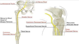
  • 45. COMMON PERONEAL NERVE • • • • ROOT VALUE = Dorsal divisions of Ventral rami of L4, L5, S1, S2. • COURSE :-. This is smaller terminal branch of the SCIATIC NERVE • It lies in the same superficial plane as the tibial nerve • . It extends from the superior angle of the fossa to the • lateral angle, along the medial border of the biceps femoris. • Continuing downwards and forwards, • it winds round the posterolateral aspect of the neck of the fibula • , pierces the peroneus longus, and divides into the superficial. • and deep peroneal nerve
  • 47. BRANCHES • [1. ] Cutaneous branches are two . (A) Lateral cutaneous nerve of the calf - descends to supply the skin of the upper two -thirds of the lateral side of the leg (B). Sural communicating nerve - arises in the upper part of the Foss. It runs on the posterolateral aspect of calf and joins the sural nerve.
  • 48. (2). ARTICULAR BRANCHES: (A). SUPERIOR LATERAL GENICULAR NERVE – accompanies the artery of the same name and lies above the lateral femoral condyle. (B). INFERIOR LATERAL GENECULAR NERVE also runs with the artery of the same name to the lateral aspect of knee joint above the head of fibula. (C). RECURRENT GENICULAR NERVE - arises where common peroneal . nerve divides into superficial and deep . peroneal nerves. It ascends anterior to the knee joint . and supplies tibialis anterior muscle in addition to the . knee joint
  • 49. • (3). Muscular branches -. • do not arise from this nerve. • However, it may give a branch to the • short head of biceps femoris..
  • 50. POSTERIOR CUTENWOUS NERVE OF THIGH • It is a content of the upper half of the popliteal fossa. • It pierces the deep fascia about the middle of the fossa, • and supplies the skin up to the middle of the back of • the Leg. GENICULAR BRANCH OF OBTURATOR NERVE This is the continuation of the posterior division of the obturator nerve. It runs on the posterior surface of the popliteal artery, pierces the oblique popliteal ligament, and supplies the capsule of the knee joint . POPLETEAL LYMPH NODES . These lie deep to the deep fascia near the termination of the small saphenous vein. They receive afferents from lateral part of sole, both superficial and deep parts Of back of leg and knee joint. The efferents end in deep inguinal lymph nodes.
  • 51. CLINICAL ANATOMY • The common peroneal nerve can be palpated against the posterolateral side of the neck of the fibula. • It may be injured in this area. It is the most frequently • injured nerve in the lower limb. • This nerve is relatively unprotected. • It may get entrapped between the attachments of peroneus • longus to the head and shaft of fibula. • Patients present “foot drop” which is usually painless. • There is weakness of dorsiflexion of ankle and of eversion • of the foot. • Inversion and plantar flexion are normal and ankle jerk is
  • 52. • Popliteal lymph nodes get enlarged in infection on lateral side of sole/foot. These are lying along the short saphenous vein. • Short saphenous vein pierces • deep fascia to drain into popliteal vein
  • 53. • Guidence by, 1. Gajendra Sir 2. Jignesh sir • Presented by, 1. Chauhan Darshna 2. Hadiyal Pooja 3. Hasti Vadariya 4. Seema Mer 5. Jay Parmar 6. Manas Joshi 7. Mahesh Vaja 8. Kamaliya Dinesh 9. Gangawala Dev