3. COMPLEX ODONTOGENIC INFECTIONS
-Natural history of progression of odontogenic infections
-Fascial spaces: fascia lined tissue compartments filled with
loose, areolar connective tissue that can become inflamed when
invaded by microorganisms
-Potential Spaces??
4. SPACES INVOLVED IN ODONTOGENIC INFECTIONS
PRIMARY SPACES
• Maxillary
Palatal
Buccal
Vestibular
• Mandibular spaces
Body of mandible
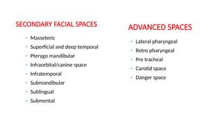
5. SECONDARY FACIAL SPACES
• Masseteric
• Superficial and deep temporal
• Pterygo mandibular
• Infraorbital/canine space
• Infratemporal
• Submandibular
• Sublingual
• Submental
• Lateral pharyngeal
• Retro pharyngeal
• Pre tracheal
• Carotid space
• Danger space
ADVANCED SPACES
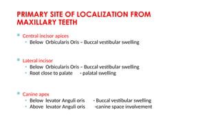
12. INFRAORBITAL/CANINE SPACE
– Thin potential space
– Between levator anguli oris & levator labii
superioris
– From maxillary canine
– Erodes superior to the origin of levator
anguli oris and below the origin of the
levator labii superioris
– Swelling obliterates the nasolabial fold
14. BUCCAL SPACE
– Bounded by overlying skin of the face on the lateral
aspect and the buccinator muscle on the medial
aspect
– From either the maxillary or mandibular teeth; most
commonly maxillary molars followed by mandibular
molars
– Infection erodes superior (or inferior) to the
attachment of the buccinator muscle
– Swelling below the zygomatic arch and above the
lower border of the mandible (Both zygomatic arch
and lower border of the mandible are palpable in
buccal space infections)
17. CAVERNOUS SINUS THROMBOSIS
(CST)
Anterior route
Erosion of infraorbital vein in infraorbital space
OR
Inferior ophthalmic vein in the sinuses
follow common ophthalmic vein to CS
Posterior route
Through infratemporal space
contains pterygoid plexus which
communicates with CS through emissary veins
20. INFECTIONS ARISING FROM MANDIBULAR TEETH
-Space of body of mandible
-Perimandibular spaces
Submandibular
Sublingual
Submental
-Masticator space
Submasseteric
Pterygomandibular
Superficial temporal
Deep tempral
21. SUBLINGUAL & SUBMANDIBULAR
SPACES
– Level of lingual perforation determinant of whether the
infection will be sublingual or submandibular
SUBLINGUAL SPACE
• Between FOM and mylohyoid
• Posterior border is open communicates freely with
the submandibular space and secondary spaces of the
mandible
• Intra oral swelling below the tongue on the affected
side usually becomes bilateral and the tongue
becomes elevated
23. SUBMANDIBULAR SPACE
• Between mylohyoid and overlying skin
and superficial fascia
• Posterior boundary communicates with
the secondary spaces
• Swelling between inferior border of
mandible, digastric and hyoid
24. LUDWIG’S ANGINA
A massive, firm cellulitis affecting simultaneously
submental, sublingual and sub mandibular spaces
bilaterally
25. SIGNS & SYMPTOMS
Facial Swelling – (Bilateral)
• -Massive, Firm,Brawny
• -May extend upto clavicles
Intra oral swelling
- Sublingual tissues
-Distension - floor of mouth
-Tongue displacement/ protrusion
-Trismus, drooling of saliva, and dysphagia, dyspnea
-Severe anxiety, disability to swallow and maintain an airway,difficult speech
Signs of Toxemia
• -High grade fever
• -Progressive dyspnoea / Cyanosis
Blood gases assessment
• -Edema glottis- Respiratory obstruction
• -Fatal within 12 to 24hrs ( Special attention to maintenance of airway)
26. TREATMENT
Admission in hospital
Secure Airway
Incision and drainage
Remove the cause
Aggressive Antibiotic therapy
27. INCISION AND DRAINAGE
Anesthesia
• -Awaked endotracheal intubation with fiberoptic laryngoscope
• -I/v analgesia + local analgesia
• -Naso pharyngeal airway/ tracheostomy set – ready
I & D
• -Separate drainage of all three spaces
• -Sublingual space drained through intra oral approach at base
of alveolar process in lingual sulcus.
• -Submandibular submental space extraoral
28. SUB MASSETERIC SPACE
– Between lateral surface of mandible and
the medial boundary of the masseter
– Most commonly from buccal space or soft
tissue infection from lower 3rd
molar
– Area overlying the ramus and angle of jaw
becomes swollen
– Moderate – severe trismus caused by
inflammation of masseter
30. PTERYGOMANDIBULAR SPACE
– Medial to mandible and lateral to medial
pterygoid muscle
– Space in which LA solution is injected
when performing an IAN block
– Primarily from sublingual or
submandibular spaces; sometimes also
from infected needles
– Little or no facial swelling patient has
significant trismus
– Trismus without swelling is a valuable
diagnostic clue
31. TEMPORAL SPACES
– Posterior and superior to masseteric and
pterygomandibular spaces
– Two portions division by temporalis muscle
• Superficial portion; between temporalis and temporalis
fascia
• Deep portion; between temporalis and temporal bone
– Involve usually only in severe infections
– Swelling in the temporal area,
superior to zygomatic arch and
posterior to lateral orbital rim
32. LATERAL PHARYNGEAL SPACE
BOUNDARIES
Anterior –superior & middle pharyngeal constrictors
Posterior – carotid sheath and scalene fascia
Superior – base of skull
Inferior – hyoid bone
Medial – pharyngeal constrictors and retropharyngeal fascia
Lateral – medial pterygoid muscle
Compartments
Anterior compartment
Posterior compartment
#17:Involvement of cavernous sinus by odontogenic infection
#18:-Papilledema. Edema of the optic disk (papilla), most commonly due to increased ICP, malignant hypertension, or thrombosis of the central retinal vein.
-a picket fence fever, spiking in the afternoon or early evening, then returning to normal.
Kernig’s sign. In dorsal decubitus, the patient can easily and completely extend the leg; in the sitting posture or when lying with the thigh flexed upon the abdomen the leg cannot be completely extended; it is a sign of meningitis.
Brudzinski’s sign. 1. In meningitis, flexion of the neck usually results in flexion of the hip and knee. 2. In meningitis, when passive flexion of the lower limb on one side is made, a similar movement will be seen in the opposite limb; called also contralateral sign.
Biot’s respiration. Breathing characterized by irregular periods of apnoea alternating with periods in which four or five breaths of identical breath are taken; seen in patients with increased ICP.
#25:May progress with alarming speed upper airway obstruction that often leads to death…Vigorous I & D procedures needed with aggressive antibiotic therapy.