L11-12.DISORDERS of the REPRODUCTIVE SYSTEM.pptx
Download as PPTX, PDF0 likes27 views

L11-12.DISORDERS of the REPRODUCTIVE SYSTEM.pptx


L11-12.DISORDERS of the REPRODUCTIVE SYSTEM.pptx
- 1. Reproductive Endocrinology ASSISTANT PROFESSOR DR BILAL NATIQ NUAMAN CONSULTANT ENDOCRINOLOGIST Al-Iraqia Medical College 2025
- 5. Male Reproductive Endocrinology In the male, the testis serves two principal functions: synthesis of testosterone by the interstitial Leydig cells under the control of luteinising hormone (LH), and spermatogenesis by Sertoli cells under the control of follicle-stimulating hormone (FSH) (but also requiring adequate testosterone). Negative feedback suppression of LH is mediated principally by testosterone, while secretion of another hormone produced by the testis, inhibin, suppresses FSH. The axis can be assessed easily by a random blood sample for testosterone, LH and FSH. Testosterone levels are higher in the morning and therefore, if testosterone is marginally low, sampling should be repeated with the patient fasted at 0900 hrs. Testicular function can also be tested by semen analysis.
- 8. A Prader orchidometer is a more reproducibly accurate method. The Prader orchidometer consists of a series of plastic ellipsoids ranging in volume from 1 to 25 mL. Each testis is compared with the appropriate ellipsoid. Adults normally have a testicular volume more than or equal to 15 mL by this method A normal testicular volume measured by ultrasound is more than 10 mL.
- 9. Female Reproductive Endocrinology In the female, physiology varies during the normal menstrual cycle. FSH stimulates growth and development of ovarian follicles during the first 14 days after the menses. This leads to a gradual increase in estradiol production from granulosa cells, which initially suppresses FSH secretion (negative feedback) but then, above a certain level, stimulates an increase in both the frequency and amplitude of gonadotrophin-releasing hormone (GnRH) pulses, resulting in a marked increase in LH secretion (positive feedback). The mid-cycle ‘surge’ of LH induces ovulation. After release of the ovum, the follicle differentiates into a corpus luteum, which secretes progesterone.
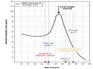
- 14. Delayed puberty Puberty is considered to be delayed if the onset of the physical features of sexual maturation has not occurred by a chronological age that is 2.5 standard deviations (SD) above the national average by the age of 14 in boys and 13 in girls.
- 16. Clinical assessment The key issue is to determine whether the delay in puberty is simply because the ‘clock is running slow’ (constitutional delay of puberty) or because there is pathology in the hypothalamus/pituitary (hypogonadotrophic hypogonadism) or the gonads (hypergonadotrophic hypogonadism). A general history and physical examination should be performed with particular reference to previous or current medical disorders, social circumstances and family history.
- 19. Body proportions, sense of smell and pubertal stage should be carefully documented and, in boys, the presence or absence of testes in the scrotum noted. Current weight and height may be plotted on centile charts, along with parental heights. Previous growth measurements in childhood, which can usually be obtained from health records, are extremely useful. Healthy growth usually follows a centile. Usually, children with constitutional delay have always been small but have maintained a normal growth velocity that is appropriate for bone age.
- 20. Constitutional delay of growth and puberty This is the most common cause of delayed puberty, but is a much more frequent explanation for lack of pubertal development in boys than in girls. Affected children are healthy and have usually been more than 2 SD below the mean height for their age throughout childhood. There is often a history of delayed puberty in siblings or parents. Since sex steroids are essential for fusion of the epiphyses, ‘bone age’ can be estimated by X-rays of epiphyses, usually in the wrist and hand; in constitutional delay, bone age is lower than chronological age. Constitutional delay of puberty should be considered as a normal variant, as puberty will commence spontaneously. However, affected children can experience significant psychological distress because of their lack of physical
- 21. Management Puberty can be induced using low doses of oral oestrogen in girls (e.g. Ethinyl estradiol 2 μg daily) or testosterone in boys (testosterone gel or depot testosterone esters). Higher doses carry a risk of early fusion of epiphyses. This therapy should be given in a specialist clinic where the progress of puberty and growth can be carefully monitored. In children with constitutional delay, this ‘priming’ therapy can be discontinued when endogenous puberty is established, usually in less than a year. In children with hypogonadism, the underlying cause should be treated and reversed if possible. If hypogonadism is permanent, sex hormone doses are gradually increased during puberty and full adult replacement doses given when development is complete.
- 22. Male hypogonadism Male hypogonadism is a clinical syndrome that results from a failure of the testes to produce adequate amounts of testosterone; this syndrome is almost always associated with impaired sperm production (androgen deficiency and impairment of sperm production), or an isolated impairment of sperm production or function with normal testosterone production.
- 23. male hypogonadism may be caused by a primary disorder of the testis (primary hypogonadism); it may be secondary to a disorder of the pituitary or hypothalamus (secondary hypogonadism). The clinical presentation of patients with deficient testosterone production or action depends on the age at onset of hypogonadism. Androgen deficiency during the second to third months of fetal development results in varying degrees of ambiguity of the genitalia and male pseudohermaphroditism. If the deficiency develops during the third trimester, defects in testicular descent leading to cryptorchidism as well as micropenis may occur.
- 24. Prepubertal onset of androgen deficiency causes eunuchoidism , which is typified by a distinctive body habitus Arm span exceeds height by greater than 5 cm, and the distance from the symphysis pubis to the floor exceeds the distance from the crown of the head to the symphysis pubis by greater than 5 cm.
- 30. Before making the diagnosis of male hypogonadism, serum testosterone concentrations must be demonstrated to be low on at least two occasions on blood samples obtained in the early morning. In addition, measurement of serum testosterone during the fasting state reduces the variability and results in higher serum testosterone concentrations.
- 31. The primary use of basal FSH and LH concentrations is to distinguish between primary (hypergonadotropic) hypogonadism (both gonadotropins are elevated) , and secondary (hypogonadotropic) hypogonadism, in which the gonadotropins are low or inappropriately normal in the presence of decreased androgen production. In primary hypogonadism, serum FSH concentrations are always higher than LH concentrations because the decreased negative feedback from inhibin B and sex steroids has greater effects on FSH secretion than the decreased negative feedback of sex steroids on LH secretion.
- 32. Elevated gonadotropin concentrations indicate primary hypogonadism due to testicular dysfunction. Serum karyotyping is useful to diagnose Klinefelter syndrome although this diagnosis may often be made clinically in males over age 16 with very small testes and biochemical evidence of primary hypogonadism.
- 33. Klinefelter syndrome is the most common genetic cause of male hypogonadism, occurring in one of 600 male births. An extra X chromosome is present in about 0.2% of male conceptions Patients with an XXY genotype often have classic Klinefelter syndrome; those with an XXXY, XXXXY, or XXYY genotype or with XXY/chromosomal mosaicism may have milder, variant forms of the syndrome.
- 34. Clinical features The diagnosis of Klinefelter syndrome is typically made in adolescents who have presented with gynaecomastia and failure to progress normally through puberty. Affected individuals usually have small, firm testes. Tall stature is apparent from early childhood, reflecting characteristically long leg length associated with 47,XXY, and may be exacerbated by androgen deficiency with lack of epiphyseal closure in puberty. Other clinical features may include learning difficulties and behavioral disorders, as well as an increased risk of breast cancer and type 2 diabetes in later life. The spectrum of clinical features is wide and some individuals, especially those with 46,XY/47,XXY mosaicism, may pass through puberty normally and be identified only during investigation for infertility.
- 35. Klinefelter syndrome is characterized by a compensatory rise in serum FSH and LH concentrations. The elevated LH concentrations stimulate aromatization of testosterone and testosterone precursor, resulting in increased production of estradiol and estradiol precursors. The relatively high estradiol : testosterone ratio causes variable degrees of gynecomastia in patients with Klinefelter syndrome.
- 36. Diagnosis and management Klinefelter syndrome is suggested by the typical phenotype in a patient with hypergonadotrophic hypogonadism and can be confirmed by karyotype analysis. Individuals with clinical and biochemical evidence of androgen deficiency require androgen replacement . There are reports of successful pregnancy occurring following ICSI therapy where spermatocytes have been retrieved from the gonads of men with Klinefelter syndrome.
- 38. GYNECOMASTIA Gynecomastia is an overdevelopment or enlargement of the breast tissue in men or boys. The breasts become larger. They often grow unevenly. It is often caused by changes in levels of the female hormone (estrogen) and the male hormone (testosterone).
- 39. The most important elements of the physical examination are breast and testicular examinations. The clinician must distinguish between pseudogynecomastia (fat tissue) and true gynecomastia. Pinching the tissue between thumb and index finger or between two hands allows the examiner to feel for an edge that represents the interface between normal tissue and rubbery glandular breast tissue. The examiner can often "flip the edge" of this interface so that the discoid breast tissue can be moved up and down or in and out of the plane of the surrounding tissue. Comparison of the consistency with fat tissue in the abdomen is useful. Gynecomastia is considered significant in adult men by some experts when there is more than or equal to 2 cm of palpable breast tissue.
- 40. The testicles should be examined for size, consistency, and the presence of a mass. A mass might represent a hormone-producing tumor. In addition to a careful examination of the breasts and testes, the clinician should examine for signs of hyperthyroidism, Cushing syndrome, and acromegaly.
- 42. Investigations If a clinical distinction between gynaecomastia and adipose tissue cannot be made, then ultrasonography or mammography is required. A random blood sample should be taken for testosterone, LH, FSH, estradiol, prolactin and hCG. Elevated oestrogen concentrations are found in testicular tumours and hCG-producing neoplasms.
- 44. Management An adolescent with gynaecomastia who is progressing normally through puberty may be reassured that the gynaecomastia will usually resolve once development is complete. If puberty does not proceed normally, then there may be an underlying abnormality that requires investigation. Gynaecomastia may cause significant psychological distress, especially in adolescent boys, and surgical excision may be justified for cosmetic reasons. Androgen replacement will usually improve gynaecomastia in hypogonadal males and any other identifiable underlying cause should be addressed if possible.
- 45. The underlying disease should be corrected if possible, and offending drugs should be discontinued. Selective estrogen receptor modulators, such as tamoxifen or raloxifene, have been found useful in relieving pain and reversing gynecomastia in some patients. Although it would be expected that reduction of estradiol production by inhibition of aromatase would be effective for gynecomastia, the largest aromatase study to date demonstrated no benefit for anastrazole compared to placebo. Short-term administration of tamoxifen (10-20 mg daily for 3-6 months) may be useful in boys with significantly symptomatic pubertal gynecomastia or men with recent onset of very symptomatic idiopathic gynecomastia. Tamoxifen also effectively prevents the development of gynecomastia in many men who are starting therapy for prostate cancer with antiandrogens. Medical therapy is
- 46. Amenorrhoea Primary amenorrhea may be diagnosed in a female who has never menstruated; this usually occurs as a manifestation of delayed puberty but may also be a consequence of anatomical defects of the female reproductive system, such as endometrial hypoplasia or vaginal agenesis. Secondary amenorrhea describes the cessation of menstruation in a female who has previously had periods. In non-pregnant women, secondary amenorrhea is almost invariably a consequence of either ovarian or hypothalamic/pituitary dysfunction. Premature ovarian failure (premature menopause) is defined, arbitrarily, as occurring before 40 years of age. Rarely, endometrial adhesions (Asherman syndrome) can form after uterine curettage, surgery or infection with
- 50. The most useful ‘test’ of ovarian function is a careful menstrual history: if menses are regular, measurement of gonadotrophins and estrogen is not necessary. In addition, ovulation can be confirmed by measuring plasma progesterone levels during the luteal phase (‘day 21 progesterone’).
- 51. Clinical assessment A history of galactorrhoea should be sought. Significant weight loss of any cause can cause amenorrhea by suppression of gonadotrophins. Weight gain may suggest hypothyroidism, Cushing’s syndrome (if other discriminatory features are present), or very rarely, a hypothalamic lesion. Hirsutism, obesity and long-standing irregular periods suggest polycystic ovary syndrome (PCOS). The presence of other autoimmune disease raises the possibility of autoimmune premature ovarian failure.
- 52. Investigations Pregnancy should be excluded in women of reproductive age by measuring urine or serum hCG. Serum LH, FSH, estradiol, prolactin, testosterone, T4 and TSH should be measured and, in the absence of a menstrual cycle, can be taken at any time. High concentrations of LH and FSH with low or low-normal estradiol suggest primary ovarian failure. Ovarian autoantibodies may be positive when there is an underlying autoimmune aetiology, and a karyotype should be performed in younger women to exclude mosaic Turner syndrome. Elevated LH, prolactin and testosterone levels with normal estradiol are common in PCOS. Low levels of LH, FSH and estradiol suggest hypothalamic or pituitary disease and a pituitary MRI is indicated .
- 53. There is some overlap in gonadotrophin and oestrogen concentrations between women with hypogonadotrophic hypogonadism and PCOS. If there is doubt as to the underlying cause of secondary amenorrhoea, then the response to 5 days of treatment with an oral progestogen (e.g. medroxyprogesterone acetate 10 mg twice daily) can be assessed. In women with PCOS, the progestogen will cause maturation of the endometrium and menstruation will occur a few days after the progestogen is stopped. In women with hypogonadotrophic hypogonadism, menstruation does not occur following progestogen withdrawal because the endometrium is atrophic as a result of oestrogen deficiency. If doubt persists in distinguishing oestrogen deficiency from a uterine abnormality, the capacity for menstruation can be tested with 1 month of treatment with cyclical oestrogen and progestogen (usually administered as a combined oral contraceptive pill). Assessment of bone mineral density by dual X-ray absorptiometry (DXA) may be appropriate in patients with low androgen and oestrogen levels.
- 56. Management Where possible, the underlying cause should be treated. For example, women with functional amenorrhoea due to excessive exercise and low weight should be encouraged to reduce their exercise and regain some weight. in oestrogen-deficient women, replacement therapy may be necessary to treat symptoms and/or to prevent osteoporosis. Women who have had a hysterectomy can be treated with oestrogen alone but those with a uterus should be treated with combined oestrogen/progestogen therapy, since unopposed oestrogen increases the risk of endometrial cancer. Cyclical hormone replacement therapy (HRT) regimens typically involve giving oestrogen on days 1–21 and progestogen on days 14–21 of the cycle, and this can be conveniently administered as the oral contraceptive pill. If estrogenic side-effects (Fluid retention, weight gain, hypertension and thrombosis) are a concern, then lower-dose oral or transdermal HRT may be more appropriate.
- 57. Turner syndrome Turner syndrome affects around 1 in 2500 females. It is classically associated with a 45,X karyotype but other cytogenetic abnormalities may be responsible, including mosaic forms (e.g. 45,X/46,XX or 45,X/46,XY) and partial deletions of an X chromosome. Ovarian tissue develops normally until the third month of gestation, but thereafter there is gonadal dysgenesis with accelerated degeneration of oöcytes and increased ovarian stromal fibrosis, resulting in ‘streak ovaries’. The inability of ovarian tissue to produce oestrogen results in loss o negative feedback and elevation of FSH and LH concentrations.
- 59. Diagnosis and management The diagnosis of Turner syndrome can be confirmed by karyotype analysis. Short stature, although not directly due to growth hormone deficiency, responds to high doses of growth hormone. Prophylactic gonadectomy is recommended for individuals with 45,X/46,XY mosaicism because there is an increased risk of gonadoblastoma. Pubertal development can be induced with oestrogen therapy but causes fusion of the epiphyses and cessation of growth. The timing of pubertal induction therefore needs to be carefully planned. Adults with Turner syndrome require long-term oestrogen replacement therapy and should be monitored periodically for the development of aortic root dilatation, hearing loss and other somatic complications.
- 62. Diagnosis of PCOS-The diagnosis of PCOS is typically based on clinical features (irregular menstrual cycles, acne, hirsutism), although additional information may be obtained with biochemical testing and sonographic examination. Polycystic ovaries tend to be enlarged and have been defined in multiple ways: (1) the presence of 10 or more cystic follicles that are between 2 and 8 mm in diameter and arranged along the subcapsular edge of the ovary in a string of pearls fashion; and (2) 12 or more follicles 2 to 9 mm in either ovary and/or an ovarian volume of 10 cm3 or greater.
- 63. Women with PCOS are at increased risk of glucose intolerance and some authorities recommend screening for type 2 diabetes and other cardiovascular risk factors associated with the metabolic syndrome.
- 64. Management This should be directed at the presenting complaint but all women with PCOS who are overweight should be encouraged to lose weight, as this can improve several symptoms, including menstrual irregularity, and reduces the risk of type 2 diabetes. most women with PCOS have oligomenorrhoea, with irregular, heavy menstrual periods. This may not require treatment unless fertility is desired. Metformin may restore regular ovulatory cycles in overweight women by reducing insulin resistance, although it is less effective than clomiphene at restoring fertility as measured by successful pregnancy. In women who have very few periods each year or are amenorrhoeic, the high oestrogen concentrations associated with PCOS can cause endometrial hyperplasia. Progestogens can be administered on a cyclical basis to induce regular shedding of the endometrium and a withdrawal bleed, or a progestogen-impregnated intrauterine coil can be fitted.
- 65. THANK YOU