SlideShare a Scribd company logo
Objective Structured Practical
Question (OSPE)
Subject: Ophthalmology
According to the course curriculum of
Bangladesh College of Physician &
Surgeon (BCPS)
AUTHOR:
Dr Md Anisur Rahman Anjum.
MBBS (Dhaka Medical College). DO (Dhaka
University) FCPS (EYE)
Associate Professor
National Institute of Ophthalmology
Dhaka, Bangladesh.
Chamber: Mojibunnessa Eye Hospital
House: 18 Road: 6. Dhanmondi, Dhaka, 1205.
Bangladesh.
Email: anjumk38dmc@gmail.com
Cell: 01711-832397
7/15/2014 2anjumk38dmc@gmail.com
OSPE:1
Q:1(a)
• 1) What does the picture
show?
• 2) What is the most
likely underlying
medical condition?
Answer:1(a)
• 1) This is the right fundal FFA taken at the venous
phase. There are multiple areas of circular dots with
both hypo and hyperfluorescein.
• 2) This most likely underlying condition is diabetic
maculopathy which has been treated with
photocoagulation.
Q:1(b)
1) What is the name of
this investigation?
2) What does this picture
show?
3) What is your
diagnosis?
Answer: 1(b)
1) This is the Fluorescence angiography of the
right fundus taken at the venous phase.
2) There are multiple spots of fluorescence (dot
& blot haemorrhage).
3) The patient has background diabetic
retinopathy.
3
Q: (1C)
1) What phase is this frame of fluorescein angiography?
2) What does the picture show? & 3) What is the diagnosis?
Answer(1c)
• 1) The late phase.
• 2) Area of hyperfluorescence with pooling. The area
with the most intense hyperfluorescence has a smoke
stack appearance. The diagnosis is CSCR.
OSPE:2
Objective structured practical question
Q:2
• The picture and fluorescein angiography are taken
from a patient complaining of distorted right vision.
• a. What phase is the fluorescein angiography?
• b. What is the diagnosis?
• c. What underlying medical condition may be present
in this patient?
ANSWER:2
 Venous phase
 Macroaneurysm. The colour picture shows circinate
exudates surrounding an area of haemorrhage. FA
shows local dilatation of the superior retinal artery
corresponding to the area of haemorrhage. The
feature is that of Macroaneurysm.
 Hypertension..
OSPE:3
Q:3
Answer the following
questions with reference
to the fluorescein
angiography.
1) What abnormality is
seen in the above
picture?
2) Suggest two possible
causes
Answer:3
1) Optic disc swelling. (The fluorescence which is
taken at venous phase shows hyper fluorescence of
the optic disc)
2)
i. Raised intracranial pressure
ii. Acute AION.
OSPE:4
Q:4
1) What imaging is
shown?
2) What does it show?
3) Name two ocular signs
which may be present?
Answer:4
1) A transverse CT scan of the brain.
2) The CT scan shows hydrocephalus.
3) 6 th nerve palsy & papillodema.
OSPE:5
This 70 year-old man suffered from progressive
left exophthalmos. His CT scan is shown as
1) What does the CT scan
show?
2) What is the most likely
diagnosis?
3) What are the
advantages of CT scan
over MRI in orbital
imaging?
Q:5
1) Hyperostosis of the left lateral portion of the
sphenoid with left proptosis.
2) Sphenoid wing meningioma.
3)
Better bony definition than MRI especially in
detecting orbital fractures and bony metastasis
Detecting metallic foreign body within the orbit
or globe (contraindicated in MRI)
Shorter running time than MRI
Less expensive than MRI
Answer:5
OSPE:6
This 70 year-old man suffered from progressive left
exophthalmos. His CT scan is shown as below
• a. What does the CT
scan show? =2
• b. What is the most
likely diagnosis? =2
• c. What are the
advantages of CT scan
over MRI in orbital
imaging? Mention 3
points =6
Q:6
Answer:6
• a. Hyperostosis of the left lateral portion of the
sphenoid with left proptosis.
• b. Sphenoid wing meningioma.
• c.
• Better bony definition than MRI especially in
detecting orbital fractures and bony metastasis
• Detecting metallic foreign body within the orbit or
globe (contraindicated in MRI)
• Shorter running time than MRI
• Less expensive than MRI
OSPE:7
ThisistheCTscanofa65year-old
womanwithunilateralproptosis.
a.WhatorientationisthisCTscan?
b.Whatabnormalitiesareshown?
c.Whatisthemostlikelydiagnosis?
d.Whatabnormalitiesmayoccurwhen
testingherocularmovement?
2.5 x 4=10
Answer:7
 a. Coronal section.
 b. Enlargement of the muscles in the right eyes
especially the inferior rectus.
 c. Right thyroid eye disease.
 d. Any of the following abnormalities may occur:
i. lid lag
ii. restricted eye movement in all directions
especially up gaze
OSPE:8
This is the CT scan of a 65 year-old woman with
unilateral (R/E) proptosis
• a. What orientation is this
CT scan?
• b. What abnormalities are
shown?
• c. What is the most likely
diagnosis?
• d. What abnormalities
may occur when testing
her ocular movement?
Answer:8
• a. Coronal section.
• b. Enlargement of the muscles in the right eyes
especially the inferior rectus.
• c. Right thyroid eye disease.
• d. Any of the following abnormalities may occur:
• lid lag
• restricted eye movement in all directions
especially up gaze
OSPE:9
Q:9
• This one year baby
was referred to the
eye clinic because of
suspected failure of
visual development.
Following
fundoscopy an
urgent CT scan was
ordered.
Q:9
• a. What does the CT scan show? =2.5
• b. What is the most likely diagnosis? =2.5
• c. Write 3 factors will determine the prognosis
of this patient? =3
• d. What is the chance of his offspring getting
the same condition? =2
Answer:9
a) The CT scan shows bilateral solid masses within the
globes with the density of the bone i.e. calcification.
b) Retinoblastoma
c)
i. Optic nerve involvement = 1
ii. Tumour size and location = 1
iii. Tumour differentiation = 1
iv. Age of the patient = 0.5
v. Secondary tumour = 0.5
d) 40%
OSPE:10
Q:10
Suppose you are working in a primary eye care hospital
at upo jilla level. A boy of 7 years old came to you
with penetrating corneal injury & iris prolapse.
Q=1
Before referring the boy at tertiary eye care centre, give
him 2 treatment.
If you are working at tertiary eye care centre, you will
manage the patient by repairing of cornea under G/A.
Q:10
• Q 2. What minimum instruments are required
to repair.=4
• Q 3. What will be your instruction to
anesthetist.=1
• Q 4. How you will decide to reposition of the
iris. Mention 2 points=3
Answer:10
ANS=1.
 Light Pad & bandage of the eye (no drop or
ointment inside the eye).
 Systemic analgesic & antibiotics.
ANS=2.
 Eye speculum.
 Barraquer needle holder
 Corneal forceps/St Martin
 2 tying forceps
Ans:10
ANS=3.
Please caring about raised IOP (avoid
Suxamethonium)
ANS=4
If iris is viable then it will reposited.
If there is a fibrinious coating over the iris
then it is sterile and can be reposited.
OSPE:11
Question
Counsel the patient
undergoing FFA.
Answer
 CHECKLIST
A. Greetings--------------------0.5
B. explanation of procedure:
i. Inj. Na Fluoride--------1.0
ii. Taking of picture----- 1.0
C. Prerequisite:
i. Dilated pupil---------1.0
ii. Renal function test—1.0
iii. Any hypersensitivity of Fluoride—1.0
Answer
D. Possible side effects:
i. Nausea/vomiting---1.0
ii. Yellow urine---------1.0
iii. Anaphylaxis /syncope—1.0
E. Talk about cost----------1.0
i. Thanks & Feedback----0.5
ii. Total-----------------------10
OSPE:11
A 50 years old lady came to you for routine eye
examination. Incidentally, it was diagnosed as
a case of POAG. How will you counseling the
lady?
Check list for observer
Done Not done
Greetings
Give idea of POAG
Rx Medical
Rx Surgical
Complications of
surgery
Fate if untreated
Follow up after
surgery
Advice
Thanks
Assessor for marking
Done Not done
Greetings 0.50
Give idea of POAG 2.50
Rx Medical 1.50
Rx Surgical 1.50
Complications of
surgery
1.00
Fate if untreated 1.50
Follow up after
surgery
1,00
Thanks 0.50
OSPE: 12
• A young patient comes to you with the
complains of uniocular sudden loss of vision.
How will you examine the patient with given
instruments- (pen torch. Snellen chart. Ishihara
chart. Ophthalmoscope.)
Check list for the observer
MARKS Done Not done
Greetings
VA
Pupil exam :
Direct
Indirect
RAPD
Colour Vision
Fundus Exam
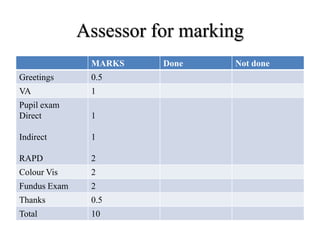
Assessor for marking
MARKS Done Not done
Greetings 0.5
VA 1
Pupil exam
Direct
Indirect
RAPD
1
1
2
Colour Vis 2
Fundus Exam 2
Thanks 0.5
Total 10
OSPE:13
Question
 Examine the simulating patient of
Keratoconus with the supplied instruments(pen
torch, ophthalmoscope, retinoscope) and
mention
1) 2 signs with pen torch.
2) 1 sign with ophthalmoscope.
3) 1 sign with retinoscope.
Answer
i. Greetings-& permission---- 1.0
ii. Rizuti reflex by torch ----1.0.
iii. Munsen sign by torch-----1.0.
iv. Oil drop reflex by ophthalmoscope—1.5.
v. Scissors reflex by retinoscope----1.5
Answer
Or if slit lamp is given then
I. -Greetings----------------0.25
II. -Positioning of patient-----0.25
III. -Positioning of slit lamp---0.5
IV. -Munsen sign( by looking down & holding the upper
lids)-------1.0
V. Diffuse illumination( see hydropic scar & volks
striae)-1.0
VI. -Oblique illumination(corneal thining/ steepening)-1.0
VII.-Fleischer ring (using cobalt blue filter)---1.0
OSPE:14
Usually BCC form a
nodule in the eyelid
but here in the picture
does not make a
nodule and grows
within the eyelid, it
induce pulling of the
eyelid.
Q:1. What is the name of this variant?
Q:2 Why this is more difficult to treat?
Q:3. Write one D/D.
Q:4. Which is the most common site in the eye?
Q:5. In which location of the eye it has the worst prognosis?
Q:6 Which will you prefer? Excisional biopsy or incisional
biopsy and why?
Answer
1) Sclerosing BCC or Morpheaform variant. = 2
2) Much more difficult to treat because its edges
are harder to define. = 2
3) Chronic blepharitis. = 1
4) Lower eyelid.= 1
5) Medial canthus. = 1
6) Excisional biopsy. Because tumours that recur
following incomplete treatment tend to be more
aggressive. = 1 + 2 = 3
OSPE:15
This is the picture of a
50 year old man who
came to you with
the painless diffuse
violet nodule in the left
lower lid. On previous
medical report you have
noted that he is
suffering from AIDS.
Question
1) What is the diagnosis of the nodule?
2) What type of tumour is this?
3) What is the causative organism?
4) In this condition, what will be the CD+4 cell
count?
1) Kaposi sarcoma.
2) This is a malignant vascular tumour
3) caused by Human Herpes Virus 8 (HHV-8)4.
4) Below 500
THANK’S A LOT FOR YOUR
PATIENCE HEARING

More Related Content

What's hot (20)

PPTX
Real pediatric refraction and spectacle power prescription
Srijana Lamichhane
 
PPTX
Dissociated vertical deviation
doc_angie Shah
 
PDF
Intrastromal Corneal Ring Segment (ICRSs)
DiyarAlzubaidy
 
PPTX
Eccentric fixation, investigation and management
Vineela Cherukuri
 
PPTX
Coat’S Disease
Dr. Jagannath Boramani
 
PPTX
Functional vision loss - Differentiating functional vision loss from malinger...
Krishna Banjade
 
PPTX
Macular hole
Md Riyaj Ali
 
PPTX
bpe.pptx
SarahKerrigan14
 
PPTX
Refractive changes after posterior segment surgeries (Dr. Nesma, TA ophthalam...
Hind Safwat
 
PPT
Diagnosis evaluation in strabismus
Jayarajini
 
PPTX
AMBLYOPIA TREATMENT STUDIES PEDIG
Sivateja Challa
 
PPTX
Lenses in ophthalmology
Reshma Peter
 
PPTX
Brown syndrome
alireza gholizade
 
PPTX
Uveitic glaucoma
SSSIHMS-PG
 
PPTX
Hess chart, diplopia chart, cover tests
Nikhil Rp
 
PPT
Choroidal coloboma
Laxmi Eye Institute
 
PPTX
Prelasik evaluation
Amr Mounir
 
PPTX
Visual rehabilitation after pediatric cataract surgery
Anuradha Chandra
 
PPT
Orthokeratology power point presentation
Digital-Marketing-Guru
 
PDF
Biometry made easy
Haitham Al Mahrouqi
 
Real pediatric refraction and spectacle power prescription
Srijana Lamichhane
 
Dissociated vertical deviation
doc_angie Shah
 
Intrastromal Corneal Ring Segment (ICRSs)
DiyarAlzubaidy
 
Eccentric fixation, investigation and management
Vineela Cherukuri
 
Coat’S Disease
Dr. Jagannath Boramani
 
Functional vision loss - Differentiating functional vision loss from malinger...
Krishna Banjade
 
Macular hole
Md Riyaj Ali
 
bpe.pptx
SarahKerrigan14
 
Refractive changes after posterior segment surgeries (Dr. Nesma, TA ophthalam...
Hind Safwat
 
Diagnosis evaluation in strabismus
Jayarajini
 
AMBLYOPIA TREATMENT STUDIES PEDIG
Sivateja Challa
 
Lenses in ophthalmology
Reshma Peter
 
Brown syndrome
alireza gholizade
 
Uveitic glaucoma
SSSIHMS-PG
 
Hess chart, diplopia chart, cover tests
Nikhil Rp
 
Choroidal coloboma
Laxmi Eye Institute
 
Prelasik evaluation
Amr Mounir
 
Visual rehabilitation after pediatric cataract surgery
Anuradha Chandra
 
Orthokeratology power point presentation
Digital-Marketing-Guru
 
Biometry made easy
Haitham Al Mahrouqi
 

Viewers also liked (20)

PPT
Ospe (objective structured practical examination)
Anisur Rahman
 
PPTX
OSPE (Ophthalmology) for FCPS, FRCOphth, MS & DO Examinee.
Anisur Rahman
 
PPTX
Objective structured practical question (ospe) for FCPS MS amd DO examinee of...
Anisur Rahman
 
PPTX
Objective structured practical question (ospe)
Anisur Rahman
 
PPTX
Workshop on ospe and osce
Bimel Kottarathil
 
PPT
Ophthalmology ospe cc
Hossein Mirzaie
 
PPTX
Cataract=4 phaco
Anisur Rahman
 
PPTX
Lecture lens
Anisur Rahman
 
PPT
Ospe tutorial
Anisur Rahman
 
PPTX
Ospe of eye
Anisur Rahman
 
PPTX
Objective structured practical question (ospe)
Anisur Rahman
 
PPTX
Eye osce
Mohd Hanafi
 
PPTX
Optics basics concepts
Anisur Rahman
 
PPTX
Tracheostomy Care
mariahenj
 
PPT
Cataract=3 ctr
Anisur Rahman
 
PDF
Ent ospe | SurgicoMed.com
Mukhdoom BaharAli
 
PPTX
Local anaesthetics
OPTOM FASLU MUHAMMED
 
PPT
Unit 1 hiv and reproductive health
David Ngogoyo
 
PPTX
Objective structured practical question (ospe)
Anisur Rahman
 
Ospe (objective structured practical examination)
Anisur Rahman
 
OSPE (Ophthalmology) for FCPS, FRCOphth, MS & DO Examinee.
Anisur Rahman
 
Objective structured practical question (ospe) for FCPS MS amd DO examinee of...
Anisur Rahman
 
Objective structured practical question (ospe)
Anisur Rahman
 
Workshop on ospe and osce
Bimel Kottarathil
 
Ophthalmology ospe cc
Hossein Mirzaie
 
Cataract=4 phaco
Anisur Rahman
 
Lecture lens
Anisur Rahman
 
Ospe tutorial
Anisur Rahman
 
Ospe of eye
Anisur Rahman
 
Objective structured practical question (ospe)
Anisur Rahman
 
Eye osce
Mohd Hanafi
 
Optics basics concepts
Anisur Rahman
 
Tracheostomy Care
mariahenj
 
Cataract=3 ctr
Anisur Rahman
 
Ent ospe | SurgicoMed.com
Mukhdoom BaharAli
 
Local anaesthetics
OPTOM FASLU MUHAMMED
 
Unit 1 hiv and reproductive health
David Ngogoyo
 
Objective structured practical question (ospe)
Anisur Rahman
 
Ad

Similar to Objective structured practical question (20)

PPTX
Ospe 25 march 2017
Anisur Rahman
 
PPTX
For slide share
Anisur Rahman
 
PPTX
Ospe 21 march 2017
Anisur Rahman
 
DOC
20 Objective Structured Clinical Examination (OSCE) for trainees.doc
Riyad Banayot
 
PPT
Management of adult cataract II.ppt
docsuleman
 
PPT
Management of adult cataract II.ppt
docsuleman
 
PPT
Management of adult cataract II.ppt
docsuleman
 
PPTX
10 short Objective Structured Clinical Examination (OSCE) Cases.pptx
Riyad Banayot
 
PDF
April 5-2013-vrics
Yesenia Castillo Salinas
 
PDF
April 5-2013-vrics
Yesenia Castillo Salinas
 
PPTX
OPHTHALMOLOGY OSCE.pptx babaBhBNhBBBbFbC
abd12medy
 
DOCX
OSCE ophthalmology examination & answers
Riyad Banayot
 
DOCX
22 Objective Structured Clinical Examination (OSCE) Ophthalmology examinatio...
Riyad Banayot
 
DOCX
Objective Structured Clinical Examination (OSCE) Ophthalmology examination.docx
Riyad Banayot
 
PPT
Osce 03ans
Nadzirah Saffian
 
DOCX
20 Multiple Choice Questions (MCQS) miscellaneous.docx
Riyad Banayot
 
PPT
Ospe eye for post graduate ophthalmic student such as FCPS. FRCOphth
Anisur Rahman
 
PDF
A Chronic Post Cataract Surgery Endophthalmitis with Suspended Intraocular Le...
CrimsonpublishersMSOR
 
PPTX
Strabismic amblyopia
drmostafahassan
 
PPTX
AUDITORY VISUAL EVOKED POTENTIAL.pptx
Dr .Priyanka Verma
 
Ospe 25 march 2017
Anisur Rahman
 
For slide share
Anisur Rahman
 
Ospe 21 march 2017
Anisur Rahman
 
20 Objective Structured Clinical Examination (OSCE) for trainees.doc
Riyad Banayot
 
Management of adult cataract II.ppt
docsuleman
 
Management of adult cataract II.ppt
docsuleman
 
Management of adult cataract II.ppt
docsuleman
 
10 short Objective Structured Clinical Examination (OSCE) Cases.pptx
Riyad Banayot
 
April 5-2013-vrics
Yesenia Castillo Salinas
 
April 5-2013-vrics
Yesenia Castillo Salinas
 
OPHTHALMOLOGY OSCE.pptx babaBhBNhBBBbFbC
abd12medy
 
OSCE ophthalmology examination & answers
Riyad Banayot
 
22 Objective Structured Clinical Examination (OSCE) Ophthalmology examinatio...
Riyad Banayot
 
Objective Structured Clinical Examination (OSCE) Ophthalmology examination.docx
Riyad Banayot
 
Osce 03ans
Nadzirah Saffian
 
20 Multiple Choice Questions (MCQS) miscellaneous.docx
Riyad Banayot
 
Ospe eye for post graduate ophthalmic student such as FCPS. FRCOphth
Anisur Rahman
 
A Chronic Post Cataract Surgery Endophthalmitis with Suspended Intraocular Le...
CrimsonpublishersMSOR
 
Strabismic amblyopia
drmostafahassan
 
AUDITORY VISUAL EVOKED POTENTIAL.pptx
Dr .Priyanka Verma
 
Ad

More from Anisur Rahman (20)

PPTX
Hypertensive retinopathy
Anisur Rahman
 
PPTX
LASER
Anisur Rahman
 
PPTX
Goldman Applanation Tonometer
Anisur Rahman
 
PPTX
Neuro-ophthalmology
Anisur Rahman
 
PPTX
Central tendency and dispersion
Anisur Rahman
 
PPTX
Ophthalmoscope direct and indirect
Anisur Rahman
 
PPTX
Neuro ophthalmology
Anisur Rahman
 
PPTX
Refractive error
Anisur Rahman
 
PPTX
04 lecture Neuro-ophthalmology
Anisur Rahman
 
PPTX
04 prism
Anisur Rahman
 
PPTX
06 lecture
Anisur Rahman
 
PPTX
03 mirror and lens
Anisur Rahman
 
PPTX
03 lecture neuro
Anisur Rahman
 
PPTX
5th lecture on research methodology
Anisur Rahman
 
PPTX
02 lecture 16 april
Anisur Rahman
 
PPTX
Second lecture neuro ophthalmology
Anisur Rahman
 
PPTX
Sample and Sampling Technique 3rd Lecture
Anisur Rahman
 
PPTX
Optics 09 april 2021
Anisur Rahman
 
PPTX
0 protocol
Anisur Rahman
 
PPTX
Ospe mbbs
Anisur Rahman
 
Hypertensive retinopathy
Anisur Rahman
 
Goldman Applanation Tonometer
Anisur Rahman
 
Neuro-ophthalmology
Anisur Rahman
 
Central tendency and dispersion
Anisur Rahman
 
Ophthalmoscope direct and indirect
Anisur Rahman
 
Neuro ophthalmology
Anisur Rahman
 
Refractive error
Anisur Rahman
 
04 lecture Neuro-ophthalmology
Anisur Rahman
 
04 prism
Anisur Rahman
 
06 lecture
Anisur Rahman
 
03 mirror and lens
Anisur Rahman
 
03 lecture neuro
Anisur Rahman
 
5th lecture on research methodology
Anisur Rahman
 
02 lecture 16 april
Anisur Rahman
 
Second lecture neuro ophthalmology
Anisur Rahman
 
Sample and Sampling Technique 3rd Lecture
Anisur Rahman
 
Optics 09 april 2021
Anisur Rahman
 
0 protocol
Anisur Rahman
 
Ospe mbbs
Anisur Rahman
 

Recently uploaded (20)

PDF
bushnell-et-al-2024-guideline-for-the-primary-prevention-of-stroke-a-guidelin...
RobertGonzalez210093
 
PPTX
Materiovigilance and Medical Device Adverse Events: A Practical Guide
Shivankan Kakkar
 
PPTX
Bill Faloon's Presentation Slides at RAADfest 2025
maximuspeto
 
PDF
Hypothyroid in practice Prof Khaled El Hadidy, Prof of internal Medicine, Hea...
Internal medicine department, faculty of Medicine Beni-Suef University Egypt
 
PDF
Development and validation of the PRISM Scale for Tomorrowmind
Yoga Tokuyoshi
 
PPTX
X-RAY PHYSICS AND IT'S PROPERTIES & FUNDAMENTALS FOR RADIOLOGY ASPIRANTS.
Shubhambharti94
 
PPTX
PCR ( Polymerase Chain Reaction) DNA sequencing .pptx
Bolan University of Medical and Health Sciences ,Quetta
 
PPTX
Decoding the Optic Disc: A Beginner’s Guide to OCT Imaging & Analysis
KafrELShiekh University
 
PPTX
4. Chest Trauma a topic of General Surgery .ppt..pptx
Bolan University of Medical and Health Sciences ,Quetta
 
PPTX
ADVANCE CARDIAC LIFE SUPPORTby Majtania.pptx
Maj Tania Bose
 
PDF
CARDIAC LIFE SUPPORT - Jagadish N. BSN RN
Jagadish N. BSN RN
 
PPTX
Code Stroke Management / Management of Acute Stroke
GODWIN SUJIN
 
PPTX
POSTPARTUM HAEMORRHAGEby Maj Taniabose.pptx
Maj Tania Bose
 
DOCX
Neuroglia - Type of nervous tissue .docx
Ayesha Fatima
 
PPT
GOOD-LABORATORY-PRACTICES-(GLP) GUIDELINES
Sushil Mahato(Pharm-D)
 
PDF
RGUHS BSc Nursing Nutrition Notes, All types of question answers are availabl...
healthscedu
 
PPTX
OBESITY and the underlying physiology.pptx
Dr. Sukriti Silwal
 
PDF
CEREBELLUM -Prof.Dr.N.Mugunthan. KMMC.pdf
Kanyakumari Medical Mission Research Center, Muttom
 
PPTX
COPD (Chronic Obstructive Pulmonary Disease) .pptx
Dr. Sukriti Silwal
 
PPTX
2025 Acute coronary syndrome Clinical Update Slide Set
desktoppc
 
bushnell-et-al-2024-guideline-for-the-primary-prevention-of-stroke-a-guidelin...
RobertGonzalez210093
 
Materiovigilance and Medical Device Adverse Events: A Practical Guide
Shivankan Kakkar
 
Bill Faloon's Presentation Slides at RAADfest 2025
maximuspeto
 
Hypothyroid in practice Prof Khaled El Hadidy, Prof of internal Medicine, Hea...
Internal medicine department, faculty of Medicine Beni-Suef University Egypt
 
Development and validation of the PRISM Scale for Tomorrowmind
Yoga Tokuyoshi
 
X-RAY PHYSICS AND IT'S PROPERTIES & FUNDAMENTALS FOR RADIOLOGY ASPIRANTS.
Shubhambharti94
 
PCR ( Polymerase Chain Reaction) DNA sequencing .pptx
Bolan University of Medical and Health Sciences ,Quetta
 
Decoding the Optic Disc: A Beginner’s Guide to OCT Imaging & Analysis
KafrELShiekh University
 
4. Chest Trauma a topic of General Surgery .ppt..pptx
Bolan University of Medical and Health Sciences ,Quetta
 
ADVANCE CARDIAC LIFE SUPPORTby Majtania.pptx
Maj Tania Bose
 
CARDIAC LIFE SUPPORT - Jagadish N. BSN RN
Jagadish N. BSN RN
 
Code Stroke Management / Management of Acute Stroke
GODWIN SUJIN
 
POSTPARTUM HAEMORRHAGEby Maj Taniabose.pptx
Maj Tania Bose
 
Neuroglia - Type of nervous tissue .docx
Ayesha Fatima
 
GOOD-LABORATORY-PRACTICES-(GLP) GUIDELINES
Sushil Mahato(Pharm-D)
 
RGUHS BSc Nursing Nutrition Notes, All types of question answers are availabl...
healthscedu
 
OBESITY and the underlying physiology.pptx
Dr. Sukriti Silwal
 
CEREBELLUM -Prof.Dr.N.Mugunthan. KMMC.pdf
Kanyakumari Medical Mission Research Center, Muttom
 
COPD (Chronic Obstructive Pulmonary Disease) .pptx
Dr. Sukriti Silwal
 
2025 Acute coronary syndrome Clinical Update Slide Set
desktoppc
 

Objective structured practical question

  • 1. Objective Structured Practical Question (OSPE) Subject: Ophthalmology According to the course curriculum of Bangladesh College of Physician & Surgeon (BCPS)
  • 2. AUTHOR: Dr Md Anisur Rahman Anjum. MBBS (Dhaka Medical College). DO (Dhaka University) FCPS (EYE) Associate Professor National Institute of Ophthalmology Dhaka, Bangladesh. Chamber: Mojibunnessa Eye Hospital House: 18 Road: 6. Dhanmondi, Dhaka, 1205. Bangladesh. Email: [email protected] Cell: 01711-832397 7/15/2014 [email protected]
  • 4. Q:1(a) • 1) What does the picture show? • 2) What is the most likely underlying medical condition?
  • 5. Answer:1(a) • 1) This is the right fundal FFA taken at the venous phase. There are multiple areas of circular dots with both hypo and hyperfluorescein. • 2) This most likely underlying condition is diabetic maculopathy which has been treated with photocoagulation.
  • 6. Q:1(b) 1) What is the name of this investigation? 2) What does this picture show? 3) What is your diagnosis?
  • 7. Answer: 1(b) 1) This is the Fluorescence angiography of the right fundus taken at the venous phase. 2) There are multiple spots of fluorescence (dot & blot haemorrhage). 3) The patient has background diabetic retinopathy.
  • 8. 3 Q: (1C) 1) What phase is this frame of fluorescein angiography? 2) What does the picture show? & 3) What is the diagnosis?
  • 9. Answer(1c) • 1) The late phase. • 2) Area of hyperfluorescence with pooling. The area with the most intense hyperfluorescence has a smoke stack appearance. The diagnosis is CSCR.
  • 12. Q:2 • The picture and fluorescein angiography are taken from a patient complaining of distorted right vision. • a. What phase is the fluorescein angiography? • b. What is the diagnosis? • c. What underlying medical condition may be present in this patient?
  • 13. ANSWER:2  Venous phase  Macroaneurysm. The colour picture shows circinate exudates surrounding an area of haemorrhage. FA shows local dilatation of the superior retinal artery corresponding to the area of haemorrhage. The feature is that of Macroaneurysm.  Hypertension..
  • 15. Q:3 Answer the following questions with reference to the fluorescein angiography. 1) What abnormality is seen in the above picture? 2) Suggest two possible causes
  • 16. Answer:3 1) Optic disc swelling. (The fluorescence which is taken at venous phase shows hyper fluorescence of the optic disc) 2) i. Raised intracranial pressure ii. Acute AION.
  • 18. Q:4 1) What imaging is shown? 2) What does it show? 3) Name two ocular signs which may be present?
  • 19. Answer:4 1) A transverse CT scan of the brain. 2) The CT scan shows hydrocephalus. 3) 6 th nerve palsy & papillodema.
  • 21. This 70 year-old man suffered from progressive left exophthalmos. His CT scan is shown as 1) What does the CT scan show? 2) What is the most likely diagnosis? 3) What are the advantages of CT scan over MRI in orbital imaging? Q:5
  • 22. 1) Hyperostosis of the left lateral portion of the sphenoid with left proptosis. 2) Sphenoid wing meningioma. 3) Better bony definition than MRI especially in detecting orbital fractures and bony metastasis Detecting metallic foreign body within the orbit or globe (contraindicated in MRI) Shorter running time than MRI Less expensive than MRI Answer:5
  • 24. This 70 year-old man suffered from progressive left exophthalmos. His CT scan is shown as below • a. What does the CT scan show? =2 • b. What is the most likely diagnosis? =2 • c. What are the advantages of CT scan over MRI in orbital imaging? Mention 3 points =6 Q:6
  • 25. Answer:6 • a. Hyperostosis of the left lateral portion of the sphenoid with left proptosis. • b. Sphenoid wing meningioma. • c. • Better bony definition than MRI especially in detecting orbital fractures and bony metastasis • Detecting metallic foreign body within the orbit or globe (contraindicated in MRI) • Shorter running time than MRI • Less expensive than MRI
  • 28. Answer:7  a. Coronal section.  b. Enlargement of the muscles in the right eyes especially the inferior rectus.  c. Right thyroid eye disease.  d. Any of the following abnormalities may occur: i. lid lag ii. restricted eye movement in all directions especially up gaze
  • 30. This is the CT scan of a 65 year-old woman with unilateral (R/E) proptosis • a. What orientation is this CT scan? • b. What abnormalities are shown? • c. What is the most likely diagnosis? • d. What abnormalities may occur when testing her ocular movement?
  • 31. Answer:8 • a. Coronal section. • b. Enlargement of the muscles in the right eyes especially the inferior rectus. • c. Right thyroid eye disease. • d. Any of the following abnormalities may occur: • lid lag • restricted eye movement in all directions especially up gaze
  • 33. Q:9 • This one year baby was referred to the eye clinic because of suspected failure of visual development. Following fundoscopy an urgent CT scan was ordered.
  • 34. Q:9 • a. What does the CT scan show? =2.5 • b. What is the most likely diagnosis? =2.5 • c. Write 3 factors will determine the prognosis of this patient? =3 • d. What is the chance of his offspring getting the same condition? =2
  • 35. Answer:9 a) The CT scan shows bilateral solid masses within the globes with the density of the bone i.e. calcification. b) Retinoblastoma c) i. Optic nerve involvement = 1 ii. Tumour size and location = 1 iii. Tumour differentiation = 1 iv. Age of the patient = 0.5 v. Secondary tumour = 0.5 d) 40%
  • 37. Q:10 Suppose you are working in a primary eye care hospital at upo jilla level. A boy of 7 years old came to you with penetrating corneal injury & iris prolapse. Q=1 Before referring the boy at tertiary eye care centre, give him 2 treatment. If you are working at tertiary eye care centre, you will manage the patient by repairing of cornea under G/A.
  • 38. Q:10 • Q 2. What minimum instruments are required to repair.=4 • Q 3. What will be your instruction to anesthetist.=1 • Q 4. How you will decide to reposition of the iris. Mention 2 points=3
  • 39. Answer:10 ANS=1.  Light Pad & bandage of the eye (no drop or ointment inside the eye).  Systemic analgesic & antibiotics. ANS=2.  Eye speculum.  Barraquer needle holder  Corneal forceps/St Martin  2 tying forceps
  • 40. Ans:10 ANS=3. Please caring about raised IOP (avoid Suxamethonium) ANS=4 If iris is viable then it will reposited. If there is a fibrinious coating over the iris then it is sterile and can be reposited.
  • 43. Answer  CHECKLIST A. Greetings--------------------0.5 B. explanation of procedure: i. Inj. Na Fluoride--------1.0 ii. Taking of picture----- 1.0 C. Prerequisite: i. Dilated pupil---------1.0 ii. Renal function test—1.0 iii. Any hypersensitivity of Fluoride—1.0
  • 44. Answer D. Possible side effects: i. Nausea/vomiting---1.0 ii. Yellow urine---------1.0 iii. Anaphylaxis /syncope—1.0 E. Talk about cost----------1.0 i. Thanks & Feedback----0.5 ii. Total-----------------------10
  • 46. A 50 years old lady came to you for routine eye examination. Incidentally, it was diagnosed as a case of POAG. How will you counseling the lady?
  • 47. Check list for observer Done Not done Greetings Give idea of POAG Rx Medical Rx Surgical Complications of surgery Fate if untreated Follow up after surgery Advice Thanks
  • 48. Assessor for marking Done Not done Greetings 0.50 Give idea of POAG 2.50 Rx Medical 1.50 Rx Surgical 1.50 Complications of surgery 1.00 Fate if untreated 1.50 Follow up after surgery 1,00 Thanks 0.50
  • 50. • A young patient comes to you with the complains of uniocular sudden loss of vision. How will you examine the patient with given instruments- (pen torch. Snellen chart. Ishihara chart. Ophthalmoscope.)
  • 51. Check list for the observer MARKS Done Not done Greetings VA Pupil exam : Direct Indirect RAPD Colour Vision Fundus Exam
  • 52. Assessor for marking MARKS Done Not done Greetings 0.5 VA 1 Pupil exam Direct Indirect RAPD 1 1 2 Colour Vis 2 Fundus Exam 2 Thanks 0.5 Total 10
  • 54. Question  Examine the simulating patient of Keratoconus with the supplied instruments(pen torch, ophthalmoscope, retinoscope) and mention 1) 2 signs with pen torch. 2) 1 sign with ophthalmoscope. 3) 1 sign with retinoscope.
  • 55. Answer i. Greetings-& permission---- 1.0 ii. Rizuti reflex by torch ----1.0. iii. Munsen sign by torch-----1.0. iv. Oil drop reflex by ophthalmoscope—1.5. v. Scissors reflex by retinoscope----1.5
  • 56. Answer Or if slit lamp is given then I. -Greetings----------------0.25 II. -Positioning of patient-----0.25 III. -Positioning of slit lamp---0.5 IV. -Munsen sign( by looking down & holding the upper lids)-------1.0 V. Diffuse illumination( see hydropic scar & volks striae)-1.0 VI. -Oblique illumination(corneal thining/ steepening)-1.0 VII.-Fleischer ring (using cobalt blue filter)---1.0
  • 58. Usually BCC form a nodule in the eyelid but here in the picture does not make a nodule and grows within the eyelid, it induce pulling of the eyelid. Q:1. What is the name of this variant? Q:2 Why this is more difficult to treat? Q:3. Write one D/D. Q:4. Which is the most common site in the eye? Q:5. In which location of the eye it has the worst prognosis? Q:6 Which will you prefer? Excisional biopsy or incisional biopsy and why?
  • 59. Answer 1) Sclerosing BCC or Morpheaform variant. = 2 2) Much more difficult to treat because its edges are harder to define. = 2 3) Chronic blepharitis. = 1 4) Lower eyelid.= 1 5) Medial canthus. = 1 6) Excisional biopsy. Because tumours that recur following incomplete treatment tend to be more aggressive. = 1 + 2 = 3
  • 61. This is the picture of a 50 year old man who came to you with the painless diffuse violet nodule in the left lower lid. On previous medical report you have noted that he is suffering from AIDS.
  • 62. Question 1) What is the diagnosis of the nodule? 2) What type of tumour is this? 3) What is the causative organism? 4) In this condition, what will be the CD+4 cell count?
  • 63. 1) Kaposi sarcoma. 2) This is a malignant vascular tumour 3) caused by Human Herpes Virus 8 (HHV-8)4. 4) Below 500
  • 64. THANK’S A LOT FOR YOUR PATIENCE HEARING