SlideShare a Scribd company logo
OSPE TUTORIALOSPE TUTORIAL
Dr Md Anisur Rahman AnjumDr Md Anisur Rahman Anjum
MBBS. DO. FCPSMBBS. DO. FCPS
Associate ProfessorAssociate Professor
National Institute of OphthalmologyNational Institute of Ophthalmology
01711-83239701711-832397
dranisurar@yahoo.comdranisurar@yahoo.com
anjumk38dmc@gmail.comanjumk38dmc@gmail.com
STATION=1STATION=1
• A young patient come to you with theA young patient come to you with the
complains of uniocular sudden loss ofcomplains of uniocular sudden loss of
vision. How will you examine the patientvision. How will you examine the patient
with given instruments- (pen torch.with given instruments- (pen torch.
Snellen chart. Ishihara chart.Snellen chart. Ishihara chart.
Ophthalmoscope.)Ophthalmoscope.)
Check list for the observerCheck list for the observer
MARKS Done Not done
Greetings
VA
Pupil exam :
Direct
Indirect
RAPD
Colour Vision
Fundus Exam
Assessor for markingAssessor for marking
MARKS Done Not done
Greetings 0.5
VA 1
Pupil exam
Direct
Indirect
RAPD
1
1
2
Colour Vis 2
Fundus Exam 2
Thanks 0.5
Total 10
STATION=2STATION=2
• This is a case of corneal injury, GiveThis is a case of corneal injury, Give
suture with 10/0 monofilament nylonsuture with 10/0 monofilament nylon
under microscope.under microscope.
Check list for the observerCheck list for the observer
Done Not Done
Wearing gloves
Adjustment
microscope:
Height
IPD
Focus
Cutting suture
Proper instrument
Procedure of
suturing
Assessor for markingAssessor for marking
CORRECT WRONG
Wearing gloves 2
Adjustment
microscope:
Height
IPD
Focus
1
1
1
Cutting suture 1
Proper instrument 1
Procedure of
suturing
3
STATION=3STATION=3
• Show the examination of LPS muscle inShow the examination of LPS muscle in
this simulated patient of congenitalthis simulated patient of congenital
ptosis.ptosis.
Check list for the observerCheck list for the observer
DONE NOT DONE
Greetings
Ask to look primary
position
Ask to look at extreme
down gaze
Hold a transparent scale
marking upper lid margin
Press thumb on eyebrows
Ask 4 look at upgaze
Fix scale
Mark the position
Thanks to patient
Assessor for markingAssessor for marking
DONE NOT DONE
Greetings 0.5
Ask to look primary
position
0.5
Ask to look at extreme
down gaze
1.5
Hold a transparent scale
marking upper lid margin
1.5
Press thumb on eyebrows 1
Ask 4 look at upgaze 1.5
Fix scale 1.5
Mark the position 1.5
Thanks to patient 0.5
STATION=4STATION=4
• You are given with Inj Vancomycin 500You are given with Inj Vancomycin 500
mg vial. Show how will you make 1 mgmg vial. Show how will you make 1 mg
in 0.1 ml.in 0.1 ml.
Check list for the observerCheck list for the observer
• Wearing of the gloves (if supplied)Wearing of the gloves (if supplied)
• Inject 10 ml D/W in the vial, so 10 ml contains 500Inject 10 ml D/W in the vial, so 10 ml contains 500
mg.mg.
• Take 1 ml from the vial, it contains 50 mg ofTake 1 ml from the vial, it contains 50 mg of
vancomycin. Add 4 ml D/W, so 5 ml contain 50 mgvancomycin. Add 4 ml D/W, so 5 ml contain 50 mg
of vancomycin.of vancomycin.
• Take 1 ml from 5 ml, now 1 ml contains 10 mg.Take 1 ml from 5 ml, now 1 ml contains 10 mg.
• Take 0.1 ml in insulin syringe which contains 1 mg ofTake 0.1 ml in insulin syringe which contains 1 mg of
vancomycin.vancomycin.
Assessor for markingAssessor for marking
• Wearing of the gloves (if supplied)Wearing of the gloves (if supplied)
• Inject 10 ml D/W in the vial, so 10 ml contains 500Inject 10 ml D/W in the vial, so 10 ml contains 500
mg.=mg.=2.52.5
• Take 1 ml from the vial, it contains 50 mg ofTake 1 ml from the vial, it contains 50 mg of
vancomycin. Add 4 ml D/W, so 5 ml contain 50 mgvancomycin. Add 4 ml D/W, so 5 ml contain 50 mg
of vancomycin.=of vancomycin.=2.52.5
• Take 1 ml from 5 ml, now 1 ml contains 10 mg.=Take 1 ml from 5 ml, now 1 ml contains 10 mg.=2.52.5
• Take 0.1 ml in insulin syringe which contains 1 mg ofTake 0.1 ml in insulin syringe which contains 1 mg of
vancomycin.=vancomycin.=2.52.5
STATION=5STATION=5
• Take relevant history from this simulatingTake relevant history from this simulating
patient of 28 years old suffering frompatient of 28 years old suffering from
transient double vision.transient double vision.
Check list for observerCheck list for observer
DONE NOT DONE
Greetings
Duration
Uni/binocular
Diurnal variation
Ocular pain
Aggravating factor
Uses of glass
Weakness of extremities
Headache
Any systematic dis
Thanks
Assessor for markingAssessor for marking
DONE NOT DONE
Greetings 0.5
Duration 1
Uni/binocular 1
Diurnal variation 1
Ocular pain 1
Aggravating factor 1
Uses of glass 1
Weakness of extremities 1
Headache 1
Any systematic dis 1
Thanks 0.5
STATION=6STATION=6
• A 50 years old lady came to you forA 50 years old lady came to you for
routine eye examination. Incidentally, itroutine eye examination. Incidentally, it
was diagnosed as a case of POAG. Howwas diagnosed as a case of POAG. How
will you counseling the lady?will you counseling the lady?
Check list for observerCheck list for observer
Done Not done
Greetings
Give idea of POAG
Rx Medical
Rx Surgical
Complications of
surgery
Fate if untreated
Follow up after
surgery
Advice
Thanks
Assessor for markingAssessor for marking
Done Not done
Greetings 0.50
Give idea of POAG 2.50
Rx Medical 1.50
Rx Surgical 1.50
Complications of
surgery
1.00
Fate if untreated 1.50
Follow up after
surgery
1,00
Thanks 0.50
STATION=7STATION=7
• Take the measurement of anteriorTake the measurement of anterior
posterior displacement of right eye.posterior displacement of right eye.
Check list for observerCheck list for observer
Greetings
Patient set up
Examinee position
Instrument placement
Bar reading adjustment
Occlusion of pt’s one
eye
Measurement of pt’s
both eyes
Thanks
Assessor for markingAssessor for marking
Done Not Done
Greetings 0.50
Patient set up 1.00
Examinee position 1.00
Instrument placement 2.00
Bar reading adjustment 2.00
Occlusion of pt’s one
eye
1.50
Measurement of pt’s
both eyes
1.50
Thanks 0.50
STATION=8
This is the chest X-ray of a patient who suffered from a
sudden uniocular visual loss. The heart is enlarged
with calcification of the left ventricle. He had a
previous history of myocardial infarction.
• What does the Chest X-ray show? Write one finding.
• What another investigation of heart you do for
diagnosis.
• What could be the cause of his sudden visual loss?
• How would you manage this patient?
1) The heart is enlarged with calcification of the left
ventricle. This is seen in left ventricular aneurysm.
2) This can be confirmed with echocardiogram.
3) What could be the cause of his sudden visual loss?
The most likely cause of his sudden visual loss is
arterial emboli arising from left ventricular
thrombus.
4How would you manage this patient?
The patient should be referred to cardiologists
for anti-coagulation treatment.
Surgical removal of the aneurysm
(aneurysmetomy) is indicated if the patient is
fit.
1) Is it T1 or T2 weighted?
2) What does the scan show?
3) What ocular signs may be present?
ANSWER
• This is an axial T1 weighted MRI scans of the
brain stem, because CSF in the ventricle is
black. There is a large tumour in the
cerebellopontine angle which pushed the
ventricle.
• Reduced corneal sensation.
• Nystagmus
• Sixth nerve palsy.
• Facial Nerve palsy.
Ospe tutorial
QUESTION
 This is the corneal topography of a patient pre-cataract
surgery.
 a. What does the picture show? = 2
 b. The patient's refraction can be corrected with
-3.00DS to 6/6.
Give a reason why he does not require cylindrical correction.
= 4
 c. A temporal clear corneal incision was performed because
the patient has deep set eye. What is the effect on:
• i. the cornea tissue = 2
ii. the cornea astigmatism = 2
ANSWER
 With the rule astigmatism.
• There is a vertical bow tie appearance of the cornea which
corresponds to that part of cornea with the steepest meridian.
 The patient may have against the rule lenticular astigmatism
which exactly neutralize the corneal astigmatism.
• The refraction of the eye is contributed by the cornea & the
lens. The astigmatism of the cornea can be neutralized by
lenticular astigmatism if its axis is opposite to that of the
cornea. For this reason, astigmatic keratometry reading during
cataract surgery should be based on the corneal topography
and not on the glass prescription.
 A temporal clear corneal incision will flatten the cornea in the
meridian of the incision.
 Because of the “coupling” the cornea will be steepened at 90
degree away. In this case, the with-the-rule-astigmatism will
increased.
STATION=8STATION=8
• A 40 year-old myopic woman is recently prescribedA 40 year-old myopic woman is recently prescribed
soft contact lenses for the first time. She returned twosoft contact lenses for the first time. She returned two
weeks later and complains that her reading visionweeks later and complains that her reading vision
is not as good as with her glasses. Retest shows heris not as good as with her glasses. Retest shows her
visual acuity to be 6/6 in both eyes with the contactvisual acuity to be 6/6 in both eyes with the contact
lenses and the lenses were of the right prescriptionlenses and the lenses were of the right prescription
and well-fitted. Why does she have problem readingand well-fitted. Why does she have problem reading
with her contact lenses but not with her glasses?with her contact lenses but not with her glasses?
ANSWERANSWER
• The patient is pre-presbyopic. Myopes requireThe patient is pre-presbyopic. Myopes require
less accommodation with glasses than contactless accommodation with glasses than contact
lenses. In addition, the prismatic effect (base-lenses. In addition, the prismatic effect (base-
in prism) offered by the concave glasses assistin prism) offered by the concave glasses assist
convergence during reading.convergence during reading.
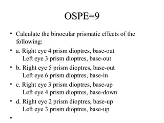
OSPE=9OSPE=9
• Calculate the binocular prismatic effects of the
following:
• a. Right eye 4 prism dioptres, base-out
Left eye 3 prism dioptres, base-out
• b. Right eye 5 prism dioptres, base-out
Left eye 6 prism dioptres, base-in
• c. Right eye 3 prism dioptres, base-up
Left eye 4 prism dioptres, base-down
• d. Right eye 2 prism dioptres, base-up
Left eye 3 prism dioptres, base-up
•
ANS OSPE=9ANS OSPE=9
• 7 prism dioptres, base-out7 prism dioptres, base-out
• 1 prism dioptre, base-in1 prism dioptre, base-in
• 7 prism dioptre, base-down7 prism dioptre, base-down
• 1 prism dioptre, base-up1 prism dioptre, base-up

More Related Content

What's hot (20)

PPTX
OSPE (Ophthalmology) for FCPS, FRCOphth, MS & DO Examinee.
Anisur Rahman
 
PPTX
Objective structured practical question (ospe)
Anisur Rahman
 
PPTX
Cataract=4 phaco
Anisur Rahman
 
PPTX
Dr screening training for nurses 7-detecting retinal disease
Riyad Banayot
 
DOCX
20 ophthalmology clinical cases
Riyad Banayot
 
PPTX
Optometry's Role in Laser Vision Correction
coakleylincoln
 
PPT
Ill-sustained accommodation
Noor Munirah Aab
 
PPTX
Pupil abnormalities
Ahmed Usman
 
DOC
Evaluating the optic nerve head in glaucoma
Riyad Banayot
 
PDF
Protocol for differential diagnosis of common ocular diseases
Puneet
 
PPTX
Glass Prescription
Azizul Islam
 
PPTX
Special tests for sensory and motor anomalies
Mero Eye
 
PPTX
Cataract management in children from optometrist perspective
Anis Suzanna Mohamad
 
PPT
Tips and tricks in squint: Dr. Madhu Karna
Madhu Karna
 
DOCX
Glaucoma questions BSCS 2019 2020
Riyad Banayot
 
PPTX
Glass prescription among youngs and educate elderly about
Dr.Amarendra Kumar
 
PPTX
AMBLYOPIA TREATMENT STUDIES PEDIG
Sivateja Challa
 
PPTX
Active Vision Therapy in Management of Amblyopia (healthkura.com)
Bikash Sapkota
 
PPTX
MA119 Chapter 31 ophthalmology and otolaryngology
BealCollegeOnline
 
DOCX
Ophthalmology assessment questions & answers
Riyad Banayot
 
OSPE (Ophthalmology) for FCPS, FRCOphth, MS & DO Examinee.
Anisur Rahman
 
Objective structured practical question (ospe)
Anisur Rahman
 
Cataract=4 phaco
Anisur Rahman
 
Dr screening training for nurses 7-detecting retinal disease
Riyad Banayot
 
20 ophthalmology clinical cases
Riyad Banayot
 
Optometry's Role in Laser Vision Correction
coakleylincoln
 
Ill-sustained accommodation
Noor Munirah Aab
 
Pupil abnormalities
Ahmed Usman
 
Evaluating the optic nerve head in glaucoma
Riyad Banayot
 
Protocol for differential diagnosis of common ocular diseases
Puneet
 
Glass Prescription
Azizul Islam
 
Special tests for sensory and motor anomalies
Mero Eye
 
Cataract management in children from optometrist perspective
Anis Suzanna Mohamad
 
Tips and tricks in squint: Dr. Madhu Karna
Madhu Karna
 
Glaucoma questions BSCS 2019 2020
Riyad Banayot
 
Glass prescription among youngs and educate elderly about
Dr.Amarendra Kumar
 
AMBLYOPIA TREATMENT STUDIES PEDIG
Sivateja Challa
 
Active Vision Therapy in Management of Amblyopia (healthkura.com)
Bikash Sapkota
 
MA119 Chapter 31 ophthalmology and otolaryngology
BealCollegeOnline
 
Ophthalmology assessment questions & answers
Riyad Banayot
 

Viewers also liked (20)

PPTX
Optics basics concepts
Anisur Rahman
 
PPTX
Objective structured practical question (ospe)
Anisur Rahman
 
PPTX
Lecture lens
Anisur Rahman
 
PPT
Ospe (objective structured practical examination)
Anisur Rahman
 
PPT
Cataract=3 ctr
Anisur Rahman
 
PPTX
How to write a scientific paper for publication
Anisur Rahman
 
PPT
introduction to Immunology 1st and 2nd lecture
the university of lahore
 
PPTX
Lect 1 introduction to immunology by dr. naeem
Hassan Ahmad
 
PPTX
Introduction to immunity
Society for Microbiology and Infection care
 
PPTX
Dr Md Anisur Rahman Optics basics concepts
Anisur Rahman
 
PPTX
Question study design
Anisur Rahman
 
PPTX
2 d & m mode
Ragil Rosyadi
 
PPTX
Light & optics
itutor
 
PPTX
Question study design
Anisur Rahman
 
PPTX
Mcqs for Ophthal pgs 1
Rajendraprasadachari
 
PPT
Visstereo
Hossein Mirzaie
 
PPT
OSPE REVISION PATHOLOGY BDS 2ND YEAR / 3RD YEAR MBBS
Ejaz Waris
 
PPT
MCQs for revision ophthalmology
DrAfiqahMF
 
PPT
Ophthalmology ospe cc
Hossein Mirzaie
 
PPTX
Antigen-Antibody Reactions
Dr-Yamna Zia
 
Optics basics concepts
Anisur Rahman
 
Objective structured practical question (ospe)
Anisur Rahman
 
Lecture lens
Anisur Rahman
 
Ospe (objective structured practical examination)
Anisur Rahman
 
Cataract=3 ctr
Anisur Rahman
 
How to write a scientific paper for publication
Anisur Rahman
 
introduction to Immunology 1st and 2nd lecture
the university of lahore
 
Lect 1 introduction to immunology by dr. naeem
Hassan Ahmad
 
Dr Md Anisur Rahman Optics basics concepts
Anisur Rahman
 
Question study design
Anisur Rahman
 
2 d & m mode
Ragil Rosyadi
 
Light & optics
itutor
 
Question study design
Anisur Rahman
 
Mcqs for Ophthal pgs 1
Rajendraprasadachari
 
Visstereo
Hossein Mirzaie
 
OSPE REVISION PATHOLOGY BDS 2ND YEAR / 3RD YEAR MBBS
Ejaz Waris
 
MCQs for revision ophthalmology
DrAfiqahMF
 
Ophthalmology ospe cc
Hossein Mirzaie
 
Antigen-Antibody Reactions
Dr-Yamna Zia
 
Ad

Similar to Ospe tutorial (20)

PPTX
Ospe 25 march 2017
Anisur Rahman
 
PPTX
Binocular Indirect Ophthalmoscopy
Dr. Shah Noor Hassan
 
PPTX
Binocular Indirect OPHTHALMOSCOPY
Kh.Towkir-Ul- Islam
 
PDF
21 STEPS with PRELIMINARY TESTS Optometry
AltheaJoySinceroBioc
 
PDF
Basic Eye Exam RESOURCE.pdf
ssuserad3332
 
PPTX
CLMMRH Ophthalmology Case Pres Reporting
PaulineMAntolo
 
PPTX
Presbyopia
Sirajudheen K Siru
 
PPTX
Presbyopia
Dr.Juleena Kunhimohammed
 
PDF
directophthalmoscope-180730142501 (2).pdf
anju468752
 
PDF
ilovepdf_merged.pdf
anju468752
 
PPTX
Direct ophthalmoscope
Rasika Walpitagamage
 
PPTX
EYE EXAMINATION(Linda).pptx
NeharikaKumari5
 
PDF
Eye examination.pdf
YoussefSrour5
 
PPTX
Refraction procedure by JAINULL ABEDIN OPTOMETRIST ODISHA
Jainull Abedin
 
PPTX
directophthalmoscope-180730142501 (2).pptx
anju468752
 
PPTX
Techniques of refraction is the process of calculation of glass power.
drbrijeshbhu
 
PPT
Orientation,history taking and examinatio.ppt
sultanovasits
 
PPTX
ppt on direct opthalmoscopy
manisaikoduri
 
PPTX
Indirect ophthalmoscopy
Shruti Laddha
 
PPTX
Indirect ophthalmoscopy and fundus drawing
Sonali Singh
 
Ospe 25 march 2017
Anisur Rahman
 
Binocular Indirect Ophthalmoscopy
Dr. Shah Noor Hassan
 
Binocular Indirect OPHTHALMOSCOPY
Kh.Towkir-Ul- Islam
 
21 STEPS with PRELIMINARY TESTS Optometry
AltheaJoySinceroBioc
 
Basic Eye Exam RESOURCE.pdf
ssuserad3332
 
CLMMRH Ophthalmology Case Pres Reporting
PaulineMAntolo
 
Presbyopia
Sirajudheen K Siru
 
directophthalmoscope-180730142501 (2).pdf
anju468752
 
ilovepdf_merged.pdf
anju468752
 
Direct ophthalmoscope
Rasika Walpitagamage
 
EYE EXAMINATION(Linda).pptx
NeharikaKumari5
 
Eye examination.pdf
YoussefSrour5
 
Refraction procedure by JAINULL ABEDIN OPTOMETRIST ODISHA
Jainull Abedin
 
directophthalmoscope-180730142501 (2).pptx
anju468752
 
Techniques of refraction is the process of calculation of glass power.
drbrijeshbhu
 
Orientation,history taking and examinatio.ppt
sultanovasits
 
ppt on direct opthalmoscopy
manisaikoduri
 
Indirect ophthalmoscopy
Shruti Laddha
 
Indirect ophthalmoscopy and fundus drawing
Sonali Singh
 
Ad

More from Anisur Rahman (20)

PPTX
Hypertensive retinopathy
Anisur Rahman
 
PPTX
LASER
Anisur Rahman
 
PPTX
Goldman Applanation Tonometer
Anisur Rahman
 
PPTX
Neuro-ophthalmology
Anisur Rahman
 
PPTX
Central tendency and dispersion
Anisur Rahman
 
PPTX
Ophthalmoscope direct and indirect
Anisur Rahman
 
PPTX
Neuro ophthalmology
Anisur Rahman
 
PPTX
Refractive error
Anisur Rahman
 
PPTX
04 lecture Neuro-ophthalmology
Anisur Rahman
 
PPTX
04 prism
Anisur Rahman
 
PPTX
06 lecture
Anisur Rahman
 
PPTX
03 mirror and lens
Anisur Rahman
 
PPTX
03 lecture neuro
Anisur Rahman
 
PPTX
5th lecture on research methodology
Anisur Rahman
 
PPTX
02 lecture 16 april
Anisur Rahman
 
PPTX
Second lecture neuro ophthalmology
Anisur Rahman
 
PPTX
Sample and Sampling Technique 3rd Lecture
Anisur Rahman
 
PPTX
Optics 09 april 2021
Anisur Rahman
 
PPTX
0 protocol
Anisur Rahman
 
PPTX
Ospe mbbs
Anisur Rahman
 
Hypertensive retinopathy
Anisur Rahman
 
Goldman Applanation Tonometer
Anisur Rahman
 
Neuro-ophthalmology
Anisur Rahman
 
Central tendency and dispersion
Anisur Rahman
 
Ophthalmoscope direct and indirect
Anisur Rahman
 
Neuro ophthalmology
Anisur Rahman
 
Refractive error
Anisur Rahman
 
04 lecture Neuro-ophthalmology
Anisur Rahman
 
04 prism
Anisur Rahman
 
06 lecture
Anisur Rahman
 
03 mirror and lens
Anisur Rahman
 
03 lecture neuro
Anisur Rahman
 
5th lecture on research methodology
Anisur Rahman
 
02 lecture 16 april
Anisur Rahman
 
Second lecture neuro ophthalmology
Anisur Rahman
 
Sample and Sampling Technique 3rd Lecture
Anisur Rahman
 
Optics 09 april 2021
Anisur Rahman
 
0 protocol
Anisur Rahman
 
Ospe mbbs
Anisur Rahman
 

Recently uploaded (20)

PDF
Balance and Equilibrium - The Vestibular System
MedicoseAcademics
 
PPTX
Amlapitta(Acid peptic Disease) Ayurvedic managment strategies
Dr. Nayan Mehar
 
PPTX
Cleaning validation SlideShare presentation
preethibs6
 
PPTX
PCR ( Polymerase Chain Reaction) DNA sequencing .pptx
Bolan University of Medical and Health Sciences ,Quetta
 
PDF
Performance Enhancing Drugs (PEDs) in Sports
Sreenivasa Reddy Thalla
 
PPT
6. Burns and skin grafting surgery II .ppt ·.ppt
Bolan University of Medical and Health Sciences ,Quetta
 
PPTX
UPDATE on NEWER MALARIA VACCINE.pptx
AshwaniSood12
 
PPTX
Immunity in Modern Science And ojas in Ayurveda
drnisheshmd
 
PDF
Pathophysiology of Artherosclerosis PPT.pdf
Miss. Pratiksha Ghodake
 
PPTX
Benign Paroxysmal Positional Vertigo (Bppv)
Tejalvarpe
 
PPTX
OBESITY and the underlying physiology.pptx
Dr. Sukriti Silwal
 
PPTX
Management Of Unruptured Intracranial Aneurysm.pptx
donogolo
 
PPTX
OMODELE MORENIKE PRESENTATION NAVDOC-4.pptx
Omodelemorenike
 
PPTX
Fundamentals of computer aided drug design.pptx
Onkar589550
 
PDF
Hypothyroid in practice Prof Khaled El Hadidy, Prof of internal Medicine, Hea...
Internal medicine department, faculty of Medicine Beni-Suef University Egypt
 
PPTX
THE GLUTEAL REGION ANATOMY PRESENTATION -pptx
LucasMagembe1
 
PDF
RGUHS BSc Nursing Nutrition Notes, All types of question answers are availabl...
healthscedu
 
PDF
SULCI, GYRI & FUNCTIONAL AREAS OF CEREBRUM-Prof.Dr.N.Mugunthan KMMC.pdf
Kanyakumari Medical Mission Research Center, Muttom
 
PPTX
Therapeutic-Diet-PPT. for B.sc nursing 2nd sem
Rekhanjali Gupta
 
PPT
Constructive Nature of Visual Processing
zahraaalkenani397
 
Balance and Equilibrium - The Vestibular System
MedicoseAcademics
 
Amlapitta(Acid peptic Disease) Ayurvedic managment strategies
Dr. Nayan Mehar
 
Cleaning validation SlideShare presentation
preethibs6
 
PCR ( Polymerase Chain Reaction) DNA sequencing .pptx
Bolan University of Medical and Health Sciences ,Quetta
 
Performance Enhancing Drugs (PEDs) in Sports
Sreenivasa Reddy Thalla
 
6. Burns and skin grafting surgery II .ppt ·.ppt
Bolan University of Medical and Health Sciences ,Quetta
 
UPDATE on NEWER MALARIA VACCINE.pptx
AshwaniSood12
 
Immunity in Modern Science And ojas in Ayurveda
drnisheshmd
 
Pathophysiology of Artherosclerosis PPT.pdf
Miss. Pratiksha Ghodake
 
Benign Paroxysmal Positional Vertigo (Bppv)
Tejalvarpe
 
OBESITY and the underlying physiology.pptx
Dr. Sukriti Silwal
 
Management Of Unruptured Intracranial Aneurysm.pptx
donogolo
 
OMODELE MORENIKE PRESENTATION NAVDOC-4.pptx
Omodelemorenike
 
Fundamentals of computer aided drug design.pptx
Onkar589550
 
Hypothyroid in practice Prof Khaled El Hadidy, Prof of internal Medicine, Hea...
Internal medicine department, faculty of Medicine Beni-Suef University Egypt
 
THE GLUTEAL REGION ANATOMY PRESENTATION -pptx
LucasMagembe1
 
RGUHS BSc Nursing Nutrition Notes, All types of question answers are availabl...
healthscedu
 
SULCI, GYRI & FUNCTIONAL AREAS OF CEREBRUM-Prof.Dr.N.Mugunthan KMMC.pdf
Kanyakumari Medical Mission Research Center, Muttom
 
Therapeutic-Diet-PPT. for B.sc nursing 2nd sem
Rekhanjali Gupta
 
Constructive Nature of Visual Processing
zahraaalkenani397
 

Ospe tutorial

  • 1. OSPE TUTORIALOSPE TUTORIAL Dr Md Anisur Rahman AnjumDr Md Anisur Rahman Anjum MBBS. DO. FCPSMBBS. DO. FCPS Associate ProfessorAssociate Professor National Institute of OphthalmologyNational Institute of Ophthalmology 01711-83239701711-832397 [email protected]@yahoo.com [email protected]@gmail.com
  • 2. STATION=1STATION=1 • A young patient come to you with theA young patient come to you with the complains of uniocular sudden loss ofcomplains of uniocular sudden loss of vision. How will you examine the patientvision. How will you examine the patient with given instruments- (pen torch.with given instruments- (pen torch. Snellen chart. Ishihara chart.Snellen chart. Ishihara chart. Ophthalmoscope.)Ophthalmoscope.)
  • 3. Check list for the observerCheck list for the observer MARKS Done Not done Greetings VA Pupil exam : Direct Indirect RAPD Colour Vision Fundus Exam
  • 4. Assessor for markingAssessor for marking MARKS Done Not done Greetings 0.5 VA 1 Pupil exam Direct Indirect RAPD 1 1 2 Colour Vis 2 Fundus Exam 2 Thanks 0.5 Total 10
  • 5. STATION=2STATION=2 • This is a case of corneal injury, GiveThis is a case of corneal injury, Give suture with 10/0 monofilament nylonsuture with 10/0 monofilament nylon under microscope.under microscope.
  • 6. Check list for the observerCheck list for the observer Done Not Done Wearing gloves Adjustment microscope: Height IPD Focus Cutting suture Proper instrument Procedure of suturing
  • 7. Assessor for markingAssessor for marking CORRECT WRONG Wearing gloves 2 Adjustment microscope: Height IPD Focus 1 1 1 Cutting suture 1 Proper instrument 1 Procedure of suturing 3
  • 8. STATION=3STATION=3 • Show the examination of LPS muscle inShow the examination of LPS muscle in this simulated patient of congenitalthis simulated patient of congenital ptosis.ptosis.
  • 9. Check list for the observerCheck list for the observer DONE NOT DONE Greetings Ask to look primary position Ask to look at extreme down gaze Hold a transparent scale marking upper lid margin Press thumb on eyebrows Ask 4 look at upgaze Fix scale Mark the position Thanks to patient
  • 10. Assessor for markingAssessor for marking DONE NOT DONE Greetings 0.5 Ask to look primary position 0.5 Ask to look at extreme down gaze 1.5 Hold a transparent scale marking upper lid margin 1.5 Press thumb on eyebrows 1 Ask 4 look at upgaze 1.5 Fix scale 1.5 Mark the position 1.5 Thanks to patient 0.5
  • 11. STATION=4STATION=4 • You are given with Inj Vancomycin 500You are given with Inj Vancomycin 500 mg vial. Show how will you make 1 mgmg vial. Show how will you make 1 mg in 0.1 ml.in 0.1 ml.
  • 12. Check list for the observerCheck list for the observer • Wearing of the gloves (if supplied)Wearing of the gloves (if supplied) • Inject 10 ml D/W in the vial, so 10 ml contains 500Inject 10 ml D/W in the vial, so 10 ml contains 500 mg.mg. • Take 1 ml from the vial, it contains 50 mg ofTake 1 ml from the vial, it contains 50 mg of vancomycin. Add 4 ml D/W, so 5 ml contain 50 mgvancomycin. Add 4 ml D/W, so 5 ml contain 50 mg of vancomycin.of vancomycin. • Take 1 ml from 5 ml, now 1 ml contains 10 mg.Take 1 ml from 5 ml, now 1 ml contains 10 mg. • Take 0.1 ml in insulin syringe which contains 1 mg ofTake 0.1 ml in insulin syringe which contains 1 mg of vancomycin.vancomycin.
  • 13. Assessor for markingAssessor for marking • Wearing of the gloves (if supplied)Wearing of the gloves (if supplied) • Inject 10 ml D/W in the vial, so 10 ml contains 500Inject 10 ml D/W in the vial, so 10 ml contains 500 mg.=mg.=2.52.5 • Take 1 ml from the vial, it contains 50 mg ofTake 1 ml from the vial, it contains 50 mg of vancomycin. Add 4 ml D/W, so 5 ml contain 50 mgvancomycin. Add 4 ml D/W, so 5 ml contain 50 mg of vancomycin.=of vancomycin.=2.52.5 • Take 1 ml from 5 ml, now 1 ml contains 10 mg.=Take 1 ml from 5 ml, now 1 ml contains 10 mg.=2.52.5 • Take 0.1 ml in insulin syringe which contains 1 mg ofTake 0.1 ml in insulin syringe which contains 1 mg of vancomycin.=vancomycin.=2.52.5
  • 14. STATION=5STATION=5 • Take relevant history from this simulatingTake relevant history from this simulating patient of 28 years old suffering frompatient of 28 years old suffering from transient double vision.transient double vision.
  • 15. Check list for observerCheck list for observer DONE NOT DONE Greetings Duration Uni/binocular Diurnal variation Ocular pain Aggravating factor Uses of glass Weakness of extremities Headache Any systematic dis Thanks
  • 16. Assessor for markingAssessor for marking DONE NOT DONE Greetings 0.5 Duration 1 Uni/binocular 1 Diurnal variation 1 Ocular pain 1 Aggravating factor 1 Uses of glass 1 Weakness of extremities 1 Headache 1 Any systematic dis 1 Thanks 0.5
  • 17. STATION=6STATION=6 • A 50 years old lady came to you forA 50 years old lady came to you for routine eye examination. Incidentally, itroutine eye examination. Incidentally, it was diagnosed as a case of POAG. Howwas diagnosed as a case of POAG. How will you counseling the lady?will you counseling the lady?
  • 18. Check list for observerCheck list for observer Done Not done Greetings Give idea of POAG Rx Medical Rx Surgical Complications of surgery Fate if untreated Follow up after surgery Advice Thanks
  • 19. Assessor for markingAssessor for marking Done Not done Greetings 0.50 Give idea of POAG 2.50 Rx Medical 1.50 Rx Surgical 1.50 Complications of surgery 1.00 Fate if untreated 1.50 Follow up after surgery 1,00 Thanks 0.50
  • 20. STATION=7STATION=7 • Take the measurement of anteriorTake the measurement of anterior posterior displacement of right eye.posterior displacement of right eye.
  • 21. Check list for observerCheck list for observer Greetings Patient set up Examinee position Instrument placement Bar reading adjustment Occlusion of pt’s one eye Measurement of pt’s both eyes Thanks
  • 22. Assessor for markingAssessor for marking Done Not Done Greetings 0.50 Patient set up 1.00 Examinee position 1.00 Instrument placement 2.00 Bar reading adjustment 2.00 Occlusion of pt’s one eye 1.50 Measurement of pt’s both eyes 1.50 Thanks 0.50
  • 24. This is the chest X-ray of a patient who suffered from a sudden uniocular visual loss. The heart is enlarged with calcification of the left ventricle. He had a previous history of myocardial infarction. • What does the Chest X-ray show? Write one finding. • What another investigation of heart you do for diagnosis. • What could be the cause of his sudden visual loss? • How would you manage this patient?
  • 25. 1) The heart is enlarged with calcification of the left ventricle. This is seen in left ventricular aneurysm. 2) This can be confirmed with echocardiogram. 3) What could be the cause of his sudden visual loss? The most likely cause of his sudden visual loss is arterial emboli arising from left ventricular thrombus.
  • 26. 4How would you manage this patient? The patient should be referred to cardiologists for anti-coagulation treatment. Surgical removal of the aneurysm (aneurysmetomy) is indicated if the patient is fit.
  • 27. 1) Is it T1 or T2 weighted? 2) What does the scan show? 3) What ocular signs may be present?
  • 28. ANSWER • This is an axial T1 weighted MRI scans of the brain stem, because CSF in the ventricle is black. There is a large tumour in the cerebellopontine angle which pushed the ventricle. • Reduced corneal sensation. • Nystagmus • Sixth nerve palsy. • Facial Nerve palsy.
  • 30. QUESTION  This is the corneal topography of a patient pre-cataract surgery.  a. What does the picture show? = 2  b. The patient's refraction can be corrected with -3.00DS to 6/6. Give a reason why he does not require cylindrical correction. = 4  c. A temporal clear corneal incision was performed because the patient has deep set eye. What is the effect on: • i. the cornea tissue = 2 ii. the cornea astigmatism = 2
  • 31. ANSWER  With the rule astigmatism. • There is a vertical bow tie appearance of the cornea which corresponds to that part of cornea with the steepest meridian.  The patient may have against the rule lenticular astigmatism which exactly neutralize the corneal astigmatism. • The refraction of the eye is contributed by the cornea & the lens. The astigmatism of the cornea can be neutralized by lenticular astigmatism if its axis is opposite to that of the cornea. For this reason, astigmatic keratometry reading during cataract surgery should be based on the corneal topography and not on the glass prescription.
  • 32.  A temporal clear corneal incision will flatten the cornea in the meridian of the incision.  Because of the “coupling” the cornea will be steepened at 90 degree away. In this case, the with-the-rule-astigmatism will increased.
  • 33. STATION=8STATION=8 • A 40 year-old myopic woman is recently prescribedA 40 year-old myopic woman is recently prescribed soft contact lenses for the first time. She returned twosoft contact lenses for the first time. She returned two weeks later and complains that her reading visionweeks later and complains that her reading vision is not as good as with her glasses. Retest shows heris not as good as with her glasses. Retest shows her visual acuity to be 6/6 in both eyes with the contactvisual acuity to be 6/6 in both eyes with the contact lenses and the lenses were of the right prescriptionlenses and the lenses were of the right prescription and well-fitted. Why does she have problem readingand well-fitted. Why does she have problem reading with her contact lenses but not with her glasses?with her contact lenses but not with her glasses?
  • 34. ANSWERANSWER • The patient is pre-presbyopic. Myopes requireThe patient is pre-presbyopic. Myopes require less accommodation with glasses than contactless accommodation with glasses than contact lenses. In addition, the prismatic effect (base-lenses. In addition, the prismatic effect (base- in prism) offered by the concave glasses assistin prism) offered by the concave glasses assist convergence during reading.convergence during reading.
  • 35. OSPE=9OSPE=9 • Calculate the binocular prismatic effects of the following: • a. Right eye 4 prism dioptres, base-out Left eye 3 prism dioptres, base-out • b. Right eye 5 prism dioptres, base-out Left eye 6 prism dioptres, base-in • c. Right eye 3 prism dioptres, base-up Left eye 4 prism dioptres, base-down • d. Right eye 2 prism dioptres, base-up Left eye 3 prism dioptres, base-up •
  • 36. ANS OSPE=9ANS OSPE=9 • 7 prism dioptres, base-out7 prism dioptres, base-out • 1 prism dioptre, base-in1 prism dioptre, base-in • 7 prism dioptre, base-down7 prism dioptre, base-down • 1 prism dioptre, base-up1 prism dioptre, base-up