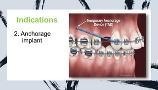
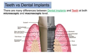
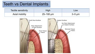
Peri-
implant
pathology
‫االمير‬ ‫عبد‬ ‫سعد‬ ‫مصطفى‬
‫العراق‬
/
‫بابل‬ ‫جامعة‬
Created By
Contents
Dental implant
A. Indications
B. Contraindication
C. Classification Of Dental Implant
D. Normal Peri-implant Mucosa
E. Peri-Implant Mucositis
F. Peri-implantitis
Dental implant
is a procedure that replaces tooth
roots with metal and screw like
posts and replaces missing teeth
with artificial teeth that look and
function much like real ones.
Indications
1. Edentulous
patient (Partially
and complete )
Indications
2. Anchorage
xc implant
Indications
3.Single tooth
loss replacement
Indications
4.Anchorage for
the maxillofacial
prosthesis
Indications
5. For rehabilitation
of congenital and
developmental
defects
Prosthodontic Rehabilitation of an Adult Patient with
Hypohidrotic Ectodermal Dysplasia
CONTRAINDICATIONS
A. General contraindications :
1. Immunologically compromised
patients
2. Cardiac diseases
3. Deficient hemostasis and blood
dyscrasias.
4. Certain psychiatric disorders
5. Recent history of orofacial
irradiation
6. Heavy smoking and alcohol abuse
B. Intraoral contraindications
1. xerostomia
2. macroglossia
3. unfavorable intermaxillary
occlusal relationship.
Classification of dental implant
1. Clinical features
• The clinically healthy gingiva and peri-implant mucosa has
a pink color and a firm consistency
• An average of 3-4 mm thickness of mucosa is ideal
Normal Soft Tissue Of Implant
2. Radiographic features :
The alveolar bone crest is usually located about 1 mm apical to a
line connecting the cemento-enamel junction of neighboring teeth.
Normal Soft Tissue Of Implant
2. Radiographic features :
The marginal termination of the bone crest is usually close to the
junction between the abutment and fixture part of the implant
system.
Normal Soft Tissue Of Implant
3. Histological features :
The mucosal tissues around
intraosseous implants form a
tightly adherent band.
This band is primarily composed of
dense collagenous lamina propria
covered by stratified squamous
keratinizing epithelium.
Normal Soft Tissue Of Implant
The junctional and barrier epithelia are about 2 mm long and the zones of
supra-alveolar connective tissues are between 1 mm and 1.5 mm high.
Both epithelia are via hemidesmosomes attached to the implant surface
Normal Soft Tissue Of Implant
• The main attachment fibers (the principal fibers) invest in the root
cementum of the tooth,
• but at the implant site the corresponding collagen fibers are nonattached
and run parallel to the implant surface, owing to the lack of cementum.
Normal Soft Tissue Of Implant
• The sulcus around an implant is lined with sulcular epithelium
that is continuous apically with the junctional epithelium
Normal Soft Tissue Of Implant
Hard Tissue
Interface
• The primary goal of
implant installation is to
achieve and maintain a
stable bone-to-implant
connection (i.e.,
osseointegration)
Hard Tissue Interface
Histologically Osseointegration : is
defined as the direct structural and
functional connection between
ordered, living bone and the surface
of a load-bearing implant without
intervening soft tissues.
Clinically, osseointegration : is the
rigid fixation of an alloplastic material
(implant) in bone with the ability to
withstand occlusal forces.
Teeth vs Dental Implants
There are many differences between Dental Implants and Teeth at both
microscopic and macroscopic level.
Teeth vs Dental Implants
Direct bone to implant
(Osseointegration)
Cementum, Alveolar bone
Periodontal ligaments
Connection
Collagen fibers ↑
fibroblast / vessels ↓
fibroblast / vessels ↑
subepithelially
Teeth vs Dental Implants
circular and
periosteogingival fibers
Dentogingival, Circular
Dentoperistal, Transseptal
fibers
Gingival fiber groups
2 groups of fibers
12 groups of fibers
Teeth vs Dental Implants
Two different sources
(the supraperiosteal vessels
and few vessels from bone)
Three different sources
(periodontal ligament space,
interdental bone, and
supraperiosteal region)
Blood supply to
surrounding gingivae
Teeth vs Dental Implants
Could be >3 mm
depending on
multiple factors
≤ 3 mm when healthy
Sulcus depth
Osseoperception
Periodontal mechanoreceptors
Proprioception
Teeth vs Dental Implants
Low
High
Tactile sensitivity
3–5 μm
25–100 μm
Axial mobility
Teeth vs Dental Implants
Crestal bone
Apical third of the root
Fulcrum when lateral
force applied
peri-implant tissues are more susceptible for inflammatory disease
than periodontal tissues. Due to the reduced :
vascularization and parallel orientation of the collagen fibers
Peri‐implant mucositis
• Peri‐implant mucositis is an inflammatory lesion of the soft
tissues surrounding an endosseous implant in the absence of
loss of supporting bone
the etiology:
The primary etiology of peri-implant mucositis
is microbial biofilm accumulation on the
implant fixture.
This biofilm is similar to that on natural
dentition,
 the inflammatory process mimic that of
gingivitis around natural teeth .
 prolonged plaque accumulation on implant
resulted in inflammation of the peri-implant
mucosa due to the establishment of an
inflammatory cell infiltrate.
 The structural difference between teeth and dental implants does not seem
to influence the host response to the bacterial insult
Peri‐implant mucositis
Risk Indicators :
To date there is some evidence to support the following as
risk indicators for the development of peri-implant mucositis:
1. Poor Oral Hygiene
2. Smoking
3. Radiation Therapy
Risk Indicators :
1. Poor Oral Hygiene
A clinical study proved there was a cause and effect relationship
between the absence of oral hygiene and plaque accumulation and
the development of peri-implant mucositis
2. Smoking
 It has been found that nicotine and its by-products can compromise the
host’s immune response, leading to reduced immune cell function
 vasoconstriction in peri-implant tissues.
 This can decrease the body’s defense
Risk Indicators :
3. Radiation Therapy
 radiation lead to changes in saliva quantity and quality, which affect
the oral microbiome and increase the risk of biofilm accumulation
around implants.
 radiation can impair the healing capacity of oral tissues, making
them more susceptible to inflammation and infection
Risk Indicators :
Potential Emerging Risk Indicators:
There is weak evidence for
1. Diabetes
2. Abutment Surface Characteristics
3. Genetics
4. Gender
5. Time Function
6. Alcohol Consumption
7. Rheumatoid Arthritis
as risk indicators for peri-implant mucositis
Diagnosis :
1. The presence of inflammatory
signs such as edema,
redness, and hyperplasia
2. The presence of BOP is
suggestive of inflammation in
peri-implant sulcus.
3. Suppuration and slight
increase in probing pocket
depth
4. Radiograph: no bone loss
Management :
Removal of plaque from the abutment/implant surface is the main goal of
treatment of peri-implant mucositis.
This goal can be achieved with
1. Patient Education 2. Oral Hygiene Instruction 3. Professional
Debridement
Patient Education
Educate each patient on the importance of adequate plaque control
This necessity in patients with history of chronic or aggressive
periodontitis and associated with tooth loss
smoking
Oral Hygiene Instruction
1.Electric vs. Manual Toothbrushes 2.Flossing vs. Interdental Brushes
3.Toothpastes 4.Mouthwashes
sonic scalers with plastic tips
Professional Debridement
titanium curettes
Professional Debridement
airflow with glycine powder
Professional Debridement
rubber cup with polishing paste
Professional Debridement
 Untreated peri‐implant mucositis may progress to
peri‐implantitis
Peri‐implantitis
• is a plaque‐associated
pathological condition occurring
in tissues around dental
implants.
• It is characterized by
inflammation in the peri‐implant
mucosa and subsequent
progressive loss of peri‐implant
bone
the etiology:
The primary etiology of peri-
implantitis is Bacterial biofilms
This biofilm is similar to that on
natural dentition,
 The response of peri-implant
tissues to the bacterial (biofilm
formation) is similar to that on
around natural teeth
both in magnitude and intensity
the etiology:
Established biofilms in implants, with increase in the inflammatory
infiltrate and loss of collagen will lead to loss of osseointegration and
implant failure.
The peri-implant lesion was considerably larger and greater apical
extension than teeth
the etiology:
Multiple other variables can influence the progression of peri-implant
disease (Table).
6. Alcohol consumption
1. History of periodontal disease
7. Implant surface
2. History of smoking
8. Occlusal overload
3. Poor oral hygiene
9. Presence of keratinized tissue
4. Diabetes
10.Iatrogenic factors
5. Genetic factors—IL-1
polymorphism
the Mobility:
ultimate result of untreated is loss of supporting bone around dental implant
As this infection develops due to plaque accumulation
bone loss occurs at the coronal aspect and progressively extends apically.
Therefore, mobility of implant
Diagnosis :
1. The presence of inflammatory
signs such as edema,
redness, and hyperplasia
2. The presence of BOP is
suggestive of inflammation in
peri-implant sulcus.
3. Suppuration and slight
increase in probing pocket
depth
4. Radiograph: bone loss is the
main diagnostic
Management :
The ultimate goal of peri-implantitis treatment is
1. Establish inflammation-free peri-implant soft tissues,
2. Eliminate all plaque
3. Prevent any further bone loss
 Treatment aims to proper oral hygiene for long-term peri-implant tissue
health
Management :
1. Nonsurgical Debridement
it shows limited efficacy when used alone for the treatment of
peri-implantitis.
This type of treatment may be suitable for esthetic areas where a
surgical approach would result in implant thread exposure and
compromised esthetics
Management :
2. Surgical Management of Peri-implantitis
A. Non-augmentative Techniques:
• Open Flap Debridement (OFD): This technique involves cleaning the
implant surface by accessing it through a flap.
• It is suitable for cases with horizontal bone loss in aesthetically
nondemanding areas.
Management :
2. Surgical Management of Peri-
implantitis
A. Non-augmentative Techniques:
• Augmentative Measures:
Recommended for peri-implantitis
sites with intrabony defects and a
minimum depth of 3 mm.
These measures can include bone
grafts or membranes
References
1. Peri-Implant Complications A
Clinical Guide to Diagnosis
and Treatment
2. Copilot (‫االصطناعي‬ ‫)الذكاء‬
THANK
for Listen

per-implant pathology (per-implanitits + Peri-Implant Mucositis)

  • 1.
    Peri- implant pathology ‫االمير‬ ‫عبد‬ ‫سعد‬‫مصطفى‬ ‫العراق‬ / ‫بابل‬ ‫جامعة‬ Created By
  • 2.
    Contents Dental implant A. Indications B.Contraindication C. Classification Of Dental Implant D. Normal Peri-implant Mucosa E. Peri-Implant Mucositis F. Peri-implantitis
  • 4.
    Dental implant is aprocedure that replaces tooth roots with metal and screw like posts and replaces missing teeth with artificial teeth that look and function much like real ones.
  • 5.
  • 6.
  • 7.
  • 8.
  • 9.
    Indications 5. For rehabilitation ofcongenital and developmental defects
  • 10.
    Prosthodontic Rehabilitation ofan Adult Patient with Hypohidrotic Ectodermal Dysplasia
  • 11.
    CONTRAINDICATIONS A. General contraindications: 1. Immunologically compromised patients 2. Cardiac diseases 3. Deficient hemostasis and blood dyscrasias. 4. Certain psychiatric disorders 5. Recent history of orofacial irradiation 6. Heavy smoking and alcohol abuse B. Intraoral contraindications 1. xerostomia 2. macroglossia 3. unfavorable intermaxillary occlusal relationship.
  • 12.
  • 16.
    1. Clinical features •The clinically healthy gingiva and peri-implant mucosa has a pink color and a firm consistency • An average of 3-4 mm thickness of mucosa is ideal Normal Soft Tissue Of Implant
  • 17.
    2. Radiographic features: The alveolar bone crest is usually located about 1 mm apical to a line connecting the cemento-enamel junction of neighboring teeth. Normal Soft Tissue Of Implant
  • 18.
    2. Radiographic features: The marginal termination of the bone crest is usually close to the junction between the abutment and fixture part of the implant system. Normal Soft Tissue Of Implant
  • 19.
    3. Histological features: The mucosal tissues around intraosseous implants form a tightly adherent band. This band is primarily composed of dense collagenous lamina propria covered by stratified squamous keratinizing epithelium. Normal Soft Tissue Of Implant
  • 20.
    The junctional andbarrier epithelia are about 2 mm long and the zones of supra-alveolar connective tissues are between 1 mm and 1.5 mm high. Both epithelia are via hemidesmosomes attached to the implant surface Normal Soft Tissue Of Implant
  • 21.
    • The mainattachment fibers (the principal fibers) invest in the root cementum of the tooth, • but at the implant site the corresponding collagen fibers are nonattached and run parallel to the implant surface, owing to the lack of cementum. Normal Soft Tissue Of Implant
  • 22.
    • The sulcusaround an implant is lined with sulcular epithelium that is continuous apically with the junctional epithelium Normal Soft Tissue Of Implant
  • 23.
    Hard Tissue Interface • Theprimary goal of implant installation is to achieve and maintain a stable bone-to-implant connection (i.e., osseointegration)
  • 24.
    Hard Tissue Interface HistologicallyOsseointegration : is defined as the direct structural and functional connection between ordered, living bone and the surface of a load-bearing implant without intervening soft tissues. Clinically, osseointegration : is the rigid fixation of an alloplastic material (implant) in bone with the ability to withstand occlusal forces.
  • 27.
    Teeth vs DentalImplants There are many differences between Dental Implants and Teeth at both microscopic and macroscopic level.
  • 28.
    Teeth vs DentalImplants Direct bone to implant (Osseointegration) Cementum, Alveolar bone Periodontal ligaments Connection Collagen fibers ↑ fibroblast / vessels ↓ fibroblast / vessels ↑ subepithelially
  • 29.
    Teeth vs DentalImplants circular and periosteogingival fibers Dentogingival, Circular Dentoperistal, Transseptal fibers Gingival fiber groups 2 groups of fibers 12 groups of fibers
  • 30.
    Teeth vs DentalImplants Two different sources (the supraperiosteal vessels and few vessels from bone) Three different sources (periodontal ligament space, interdental bone, and supraperiosteal region) Blood supply to surrounding gingivae
  • 31.
    Teeth vs DentalImplants Could be >3 mm depending on multiple factors ≤ 3 mm when healthy Sulcus depth Osseoperception Periodontal mechanoreceptors Proprioception
  • 32.
    Teeth vs DentalImplants Low High Tactile sensitivity 3–5 μm 25–100 μm Axial mobility
  • 33.
    Teeth vs DentalImplants Crestal bone Apical third of the root Fulcrum when lateral force applied
  • 34.
    peri-implant tissues aremore susceptible for inflammatory disease than periodontal tissues. Due to the reduced : vascularization and parallel orientation of the collagen fibers
  • 35.
    Peri‐implant mucositis • Peri‐implantmucositis is an inflammatory lesion of the soft tissues surrounding an endosseous implant in the absence of loss of supporting bone
  • 36.
    the etiology: The primaryetiology of peri-implant mucositis is microbial biofilm accumulation on the implant fixture. This biofilm is similar to that on natural dentition,  the inflammatory process mimic that of gingivitis around natural teeth .  prolonged plaque accumulation on implant resulted in inflammation of the peri-implant mucosa due to the establishment of an inflammatory cell infiltrate.  The structural difference between teeth and dental implants does not seem to influence the host response to the bacterial insult
  • 37.
    Peri‐implant mucositis Risk Indicators: To date there is some evidence to support the following as risk indicators for the development of peri-implant mucositis: 1. Poor Oral Hygiene 2. Smoking 3. Radiation Therapy
  • 38.
    Risk Indicators : 1.Poor Oral Hygiene A clinical study proved there was a cause and effect relationship between the absence of oral hygiene and plaque accumulation and the development of peri-implant mucositis
  • 39.
    2. Smoking  Ithas been found that nicotine and its by-products can compromise the host’s immune response, leading to reduced immune cell function  vasoconstriction in peri-implant tissues.  This can decrease the body’s defense Risk Indicators :
  • 40.
    3. Radiation Therapy radiation lead to changes in saliva quantity and quality, which affect the oral microbiome and increase the risk of biofilm accumulation around implants.  radiation can impair the healing capacity of oral tissues, making them more susceptible to inflammation and infection Risk Indicators :
  • 41.
    Potential Emerging RiskIndicators: There is weak evidence for 1. Diabetes 2. Abutment Surface Characteristics 3. Genetics 4. Gender 5. Time Function 6. Alcohol Consumption 7. Rheumatoid Arthritis as risk indicators for peri-implant mucositis
  • 42.
    Diagnosis : 1. Thepresence of inflammatory signs such as edema, redness, and hyperplasia 2. The presence of BOP is suggestive of inflammation in peri-implant sulcus. 3. Suppuration and slight increase in probing pocket depth 4. Radiograph: no bone loss
  • 43.
    Management : Removal ofplaque from the abutment/implant surface is the main goal of treatment of peri-implant mucositis. This goal can be achieved with 1. Patient Education 2. Oral Hygiene Instruction 3. Professional Debridement
  • 44.
    Patient Education Educate eachpatient on the importance of adequate plaque control This necessity in patients with history of chronic or aggressive periodontitis and associated with tooth loss smoking
  • 45.
    Oral Hygiene Instruction 1.Electricvs. Manual Toothbrushes 2.Flossing vs. Interdental Brushes 3.Toothpastes 4.Mouthwashes
  • 46.
    sonic scalers withplastic tips Professional Debridement Professional Debridement
  • 47.
  • 48.
    airflow with glycinepowder Professional Debridement Professional Debridement
  • 49.
    rubber cup withpolishing paste Professional Debridement
  • 50.
     Untreated peri‐implantmucositis may progress to peri‐implantitis
  • 51.
    Peri‐implantitis • is aplaque‐associated pathological condition occurring in tissues around dental implants. • It is characterized by inflammation in the peri‐implant mucosa and subsequent progressive loss of peri‐implant bone
  • 52.
    the etiology: The primaryetiology of peri- implantitis is Bacterial biofilms This biofilm is similar to that on natural dentition,  The response of peri-implant tissues to the bacterial (biofilm formation) is similar to that on around natural teeth both in magnitude and intensity
  • 53.
    the etiology: Established biofilmsin implants, with increase in the inflammatory infiltrate and loss of collagen will lead to loss of osseointegration and implant failure. The peri-implant lesion was considerably larger and greater apical extension than teeth
  • 54.
    the etiology: Multiple othervariables can influence the progression of peri-implant disease (Table). 6. Alcohol consumption 1. History of periodontal disease 7. Implant surface 2. History of smoking 8. Occlusal overload 3. Poor oral hygiene 9. Presence of keratinized tissue 4. Diabetes 10.Iatrogenic factors 5. Genetic factors—IL-1 polymorphism
  • 55.
    the Mobility: ultimate resultof untreated is loss of supporting bone around dental implant As this infection develops due to plaque accumulation bone loss occurs at the coronal aspect and progressively extends apically. Therefore, mobility of implant
  • 56.
    Diagnosis : 1. Thepresence of inflammatory signs such as edema, redness, and hyperplasia 2. The presence of BOP is suggestive of inflammation in peri-implant sulcus. 3. Suppuration and slight increase in probing pocket depth 4. Radiograph: bone loss is the main diagnostic
  • 57.
    Management : The ultimategoal of peri-implantitis treatment is 1. Establish inflammation-free peri-implant soft tissues, 2. Eliminate all plaque 3. Prevent any further bone loss  Treatment aims to proper oral hygiene for long-term peri-implant tissue health
  • 58.
    Management : 1. NonsurgicalDebridement it shows limited efficacy when used alone for the treatment of peri-implantitis. This type of treatment may be suitable for esthetic areas where a surgical approach would result in implant thread exposure and compromised esthetics
  • 59.
    Management : 2. SurgicalManagement of Peri-implantitis A. Non-augmentative Techniques: • Open Flap Debridement (OFD): This technique involves cleaning the implant surface by accessing it through a flap. • It is suitable for cases with horizontal bone loss in aesthetically nondemanding areas.
  • 60.
    Management : 2. SurgicalManagement of Peri- implantitis A. Non-augmentative Techniques: • Augmentative Measures: Recommended for peri-implantitis sites with intrabony defects and a minimum depth of 3 mm. These measures can include bone grafts or membranes
  • 61.
    References 1. Peri-Implant ComplicationsA Clinical Guide to Diagnosis and Treatment 2. Copilot (‫االصطناعي‬ ‫)الذكاء‬
  • 62.