


![ [1] U. Avni, H. Greenspan, E. Konen, M. Sharon, J. Goldberger, X-ray
categorization and retrieval on the organ and pathology level, using patch-
based visual words, IEEE Trans. Med. Imaging 30 (3) (2011) 733–746
.
[2] P. Pattrapisetwong, W. Chiracharit, Automatic lung segmentation in
chest radiographs using shadow filter and multilevel thresholding, in: 2016
International Computer Science and Engineering Conference (ICSEC),
IEEE, 2016, pp. 1–6.
[3] S. Katsuragawa, K. Doi, Computer-aided diagnosis in chest
radiography, Comput. Med. Imaging Graph. 31 (4–5) (2007) 212–223.
[4] Q. Li, R.M. Nishikawa, Computer-aided Detection and Diagnosis in
Medical Imaging, Taylor & Francis, 2015.
References:](https://image.slidesharecdn.com/vivappt1-230318103440-5cbad772/85/x-RAYS-PROJECT-32-320.jpg)
![ [5] C. Qin, D. Yao, Y. Shi, Z. Song, Computer-aided detection in chest radiography based on artificial
intelligence: a survey, Biomed. Eng. Online 17 (1) (2018)
[6] A.A. El-Solh, C.-B. Hsiao, S. Goodnough, J. Serghani, B.J. Grant, Predicting active pulmonary
tuberculosis using an artificial neural network, Chest 116 (4)(1999) 968–973.
[7] O. Er, N. Yumusak, F. Temurtas, Chest diseases diagnosis using artificial neural networks, Expert Syst.
Appl. 37 (12) (2010) 7648–7655.
[8] O. Er, C. Sertkaya, F. Temurtas, A.C. Tanrikulu, A comparative study on chronic obstructive pulmonary
and pneumonia diseases diagnosis using neural networks and artificial immune system, J. Med. Syst. 33 (6)
(2009) 485–492.
[9] S. Khobragade, A. Tiwari, C. Patil, V. Narke, Automatic detection of major lung diseases using chest
radiographs and classification by feed-forward artificial neural network, in: 2016 IEEE 1st International
Conference on Power Electronics, Intelligent Control and Energy Systems (ICPEICES), IEEE, 2016,
[10] G.G.R. Schramek, D. Stoevesandt, A. Reising, J.T. Kielstein, M. Hiss, H. Kielstein,
Imaging in anatomy: a comparison of imaging techniques in embalmed human cadavers, BMC
Med. Educ. 13 (1) (2013) 143.
References:](https://image.slidesharecdn.com/vivappt1-230318103440-5cbad772/85/x-RAYS-PROJECT-33-320.jpg)

x-RAYS PROJECT
- 1. DETECTING PNEUMONIA IN CHESTX-RAYSUSING DEEP LEARNINGTECHNIQUE Project Guide: Dr.T. Jemima Jebaseeli Done by: Punithavathy S P PRK19CS1011
- 2. India accounts for 20 percent of the death worldwide caused by pneumonia Pneumonia affects the lungs and causes about 18% of all deaths in children under five years old. Chest X-rays are the most common and easiest way to detect pneumonia. X-ray images are often unclear and can be confused with other diseases that have similar features. Problem Statement
- 3. To reduce the human eye error in diagnosing the disease. To develop a deep learning framework to automatically diagnose pneumonia using chest X-ray images. To classify the result as normal cases or pneumonia cases. Objective
- 4. Machine Learning library ,sklearn used with many subordinate models such as model classifiers, and metrics etc. Deep Learning library: Keras, Pytorch etc.. Google Colab used for implementation. Data set available on open source platforms Programming Language: Python TECHNIQUES/ TECHNOLOGIES USED
- 5. The dataset is taken from kaggle. The dataset is organized into 2 folders (train, test) and contains subfolders for each image category (Pneumonia/Normal). Dataset
- 6. Methodology: IMAGEACQUISITION IMAGE PREPROCESSING SEGMENTATION FEATURE EXTRACTION NORMAL CASES PNEUMONIA CASES CNN CLASSIFICATION
- 7. Preprocessing Preprocessing Preprocessing is the removal of systematic noise from the data Image generator object which performs random rotations, shifts, flips, crops, and sheers on the image dataset. Enhancement • Image enhancement is the process of adjusting digital images so that the results are more suitable for display or further image analysis. • Enhancement remove noise, sharpen, or brighten an image, making it easier to identify key features.
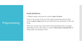
- 8. Preprocessing NOISE REMOVAL: • Digital images are prone to various types of noise. Noise is the result of errors in the image acquisition process that result in pixel values that do not reflect the true intensities of the real scene There are several ways that noise can be introduced into an image, depending on how the image is created. Electronic transmission of image data can introduce noise.
- 9. SEGMENTATION: • Image segmentation is the process of dividing an image into multiple parts. • Simplify and easier to analyze. • This is typically used to identify objects or other relevant information in digital images. segmentation:
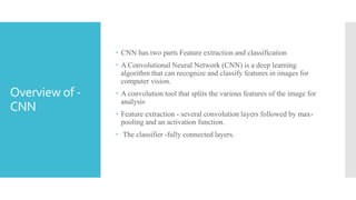
- 10. CNN has two parts Feature extraction and classification A Convolutional Neural Network (CNN) is a deep learning algorithm that can recognize and classify features in images for computer vision. A convolution tool that splits the various features of the image for analysis Feature extraction - several convolution layers followed by max- pooling and an activation function. The classifier -fully connected layers. Overview of - CNN
- 11. CNNArchitecture
- 12. Convolutional layer works by placing a filter over an array of image pixels. Convolved feature map Convolution layer
- 13. Redues the sample size Makes the process much faster Pooled feature map Two types Max pooling Average pooling Pooling Layer
- 14. CONVOLUTIO NAL NEURAL NETWORK Layers: Convolution Layer ReLu Layer Pooling Layer Fully Connected Layer
- 16. Accuracy Accuracy , precision and recall on testing set:
- 17. VGG-16
- 18. VGG-16 VGG stands for Visual Geometry Group TheVGG architecture consists of blocks, where each block is composed of 2D Convolution and Max Pooling layers. There are total of 13 convolutional layers and 3 fully connected layers inVGG16 architecture. VGG-16 is a convolutional neural network that is 16 layers deep. The network has an image input size of 224-by-224.
- 19. Architecture
- 20. VGG-16 layers
- 22. Accuracy
- 23. Mobile NetV2
- 24. Mobile net MobileNetV2 is a convolutional neural network architecture that seeks to perform well on mobile devices. It is based on an inverted residual structure where the residual connections are between the bottleneck layers The architecture of MobileNetV2 contains the initial fully convolution layer with 32 filters, followed by 19 residual bottleneck layers. ...
- 25. Architecture
- 28. Accuracy
- 29. Output Comparision CNN Model Accuracy = 90.6% VGG-16 Accuracy = 94.3% Mobile net model Accuracy = 85.00%
- 30. INFERENCE •CNN model for Pneumonia disease detection –implemented. •Mobile Net model Pneumonia Pneumonia Disease detection – implemented. VGG-16 model for Pneumonia disease detection –implemented Classification of all models –tested. VGG-16 outperformed Mobile Net model, CNN Model for pneumonia disease detection in Chest X-rays.
- 31. CONCLUSION •Pneumonia identified using Deep learning models. •Pneumonia Disease was classified using Deep learning models. •CNN model ,VGG-16 and Mobile net was used and compared for accuracy and performance. •VGG-16 model proved to produce accuracy than CNN Model and Mobile net for detecting Pneumonia disease in Chest X-rays.
- 32. [1] U. Avni, H. Greenspan, E. Konen, M. Sharon, J. Goldberger, X-ray categorization and retrieval on the organ and pathology level, using patch- based visual words, IEEE Trans. Med. Imaging 30 (3) (2011) 733–746 . [2] P. Pattrapisetwong, W. Chiracharit, Automatic lung segmentation in chest radiographs using shadow filter and multilevel thresholding, in: 2016 International Computer Science and Engineering Conference (ICSEC), IEEE, 2016, pp. 1–6. [3] S. Katsuragawa, K. Doi, Computer-aided diagnosis in chest radiography, Comput. Med. Imaging Graph. 31 (4–5) (2007) 212–223. [4] Q. Li, R.M. Nishikawa, Computer-aided Detection and Diagnosis in Medical Imaging, Taylor & Francis, 2015. References:
- 33. [5] C. Qin, D. Yao, Y. Shi, Z. Song, Computer-aided detection in chest radiography based on artificial intelligence: a survey, Biomed. Eng. Online 17 (1) (2018) [6] A.A. El-Solh, C.-B. Hsiao, S. Goodnough, J. Serghani, B.J. Grant, Predicting active pulmonary tuberculosis using an artificial neural network, Chest 116 (4)(1999) 968–973. [7] O. Er, N. Yumusak, F. Temurtas, Chest diseases diagnosis using artificial neural networks, Expert Syst. Appl. 37 (12) (2010) 7648–7655. [8] O. Er, C. Sertkaya, F. Temurtas, A.C. Tanrikulu, A comparative study on chronic obstructive pulmonary and pneumonia diseases diagnosis using neural networks and artificial immune system, J. Med. Syst. 33 (6) (2009) 485–492. [9] S. Khobragade, A. Tiwari, C. Patil, V. Narke, Automatic detection of major lung diseases using chest radiographs and classification by feed-forward artificial neural network, in: 2016 IEEE 1st International Conference on Power Electronics, Intelligent Control and Energy Systems (ICPEICES), IEEE, 2016, [10] G.G.R. Schramek, D. Stoevesandt, A. Reising, J.T. Kielstein, M. Hiss, H. Kielstein, Imaging in anatomy: a comparison of imaging techniques in embalmed human cadavers, BMC Med. Educ. 13 (1) (2013) 143. References:
- 34. Thank you…!